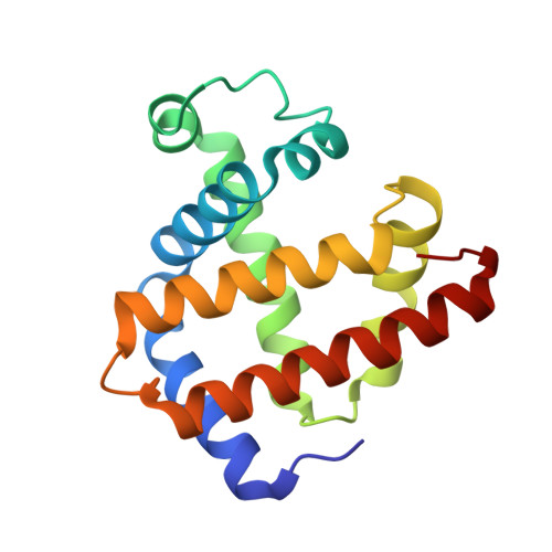
3A2G

Crystal Structure of K102C-Myoglobin conjugated with Fluorescein


Experimental Data Snapshot

  • Method: X-RAY DIFFRACTION
  • Resolution: 1.75 Å
  • R-Value Free: 0.197 
  • R-Value Work: 0.178 
  • R-Value Observed: 0.179 

Starting Model: experimental
View more details

wwPDB Validation   3D Report Full Report


Ligand Structure Quality Assessment 


This is version 1.4 of the entry. See complete history


Literature

Modification of porous protein crystals in development of biohybrid materials

Koshiyama, T.Kawaba, N.Hikage, T.Shirai, M.Miura, Y.Huang, C.-Y.Tanaka, K.Watanabe, Y.Ueno, T.

(2010) Bioconjug Chem 21: 264-269

  • DOI: https://doi.org/10.1021/bc9003052
  • Primary Citation of Related Structures:  
    3A2G

  • PubMed Abstract: 

    Protein assemblies have attracted increasing attention for construction of biohybrid materials. Protein crystals can also be regarded as solid protein assemblies. The present work demonstrates that protein crystals can be employed as porous biomaterials by site-specific modifications of the crystals of recombinant sperm whale myoglobin mutants. The myoglobin crystals of space group P6 provide hexagonal pores consisting of the building blocks of six Mb molecules, which form a pore with a diameter of 40 A. On the basis of the lattice structure of the Mb crystals, we have selected appropriate residues located on the surface of the pores for replacement with cysteine. This enables modification of the pore surface via coupling with maleimide derivatives. We have succeeded in crystallizing the modified Mb mutants, retaining the P6 lattice, and consistently aligning nanosized functional molecules such as fluorescein, eosin, and Ru(bpy)(3) into the hexagonal pores of the Mb crystals. Our strategy for site-specific modification of protein crystal pores is applicable to various protein crystals with porous structures. We believe that modified porous protein crystals will provide attractive candidates for novel solid materials in nanotechnology applications.


  • Organizational Affiliation

    Institute for Integrated Cell-Material Sciences, Funai Center, Kyoto University Katsura, Nishikyo-ku, Kyoto 615-8510, Japan.


Macromolecules
Find similar proteins by:  (by identity cutoff)  |  3D Structure
Entity ID: 1
MoleculeChains Sequence LengthOrganismDetailsImage
Myoglobin154Physeter macrocephalusMutation(s): 1 
Gene Names: MB
EC: 1.11.1 (UniProt), 1.7 (UniProt)
UniProt
Find proteins for P02185 (Physeter macrocephalus)
Explore P02185 
Go to UniProtKB:  P02185
Entity Groups  
Sequence Clusters30% Identity50% Identity70% Identity90% Identity95% Identity100% Identity
UniProt GroupP02185
Sequence Annotations
Expand
  • Reference Sequence
Small Molecules
Ligands 4 Unique
IDChains Name / Formula / InChI Key2D Diagram3D Interactions
HEM
Query on HEM

Download Ideal Coordinates CCD File 
B [auth A]PROTOPORPHYRIN IX CONTAINING FE
C34 H32 Fe N4 O4
KABFMIBPWCXCRK-RGGAHWMASA-L
LCY
Query on LCY

Download Ideal Coordinates CCD File 
G [auth A]1-methylpyrrolidine-2,5-dione
C5 H7 N O2
KYEACNNYFNZCST-UHFFFAOYSA-N
SO4
Query on SO4

Download Ideal Coordinates CCD File 
C [auth A],
D [auth A],
E [auth A]
SULFATE ION
O4 S
QAOWNCQODCNURD-UHFFFAOYSA-L
GOL
Query on GOL

Download Ideal Coordinates CCD File 
F [auth A]GLYCEROL
C3 H8 O3
PEDCQBHIVMGVHV-UHFFFAOYSA-N
Experimental Data & Validation

Experimental Data

  • Method: X-RAY DIFFRACTION
  • Resolution: 1.75 Å
  • R-Value Free: 0.197 
  • R-Value Work: 0.178 
  • R-Value Observed: 0.179 
  • Space Group: P 6
Unit Cell:
Length ( Å )Angle ( ˚ )
a = 90.409α = 90
b = 90.409β = 90
c = 45.32γ = 120
Software Package:
Software NamePurpose
REFMACrefinement
HKL-2000data collection
SCALEPACKdata scaling
MOLREPphasing

Structure Validation

View Full Validation Report


Ligand Structure Quality Assessment 


Entry History 

Deposition Data

Revision History  (Full details and data files)

  • Version 1.0: 2010-03-02
    Type: Initial release
  • Version 1.1: 2011-07-13
    Changes: Version format compliance
  • Version 1.2: 2021-11-10
    Changes: Database references, Derived calculations
  • Version 1.3: 2023-11-01
    Changes: Data collection, Refinement description
  • Version 1.4: 2024-10-30
    Changes: Structure summary