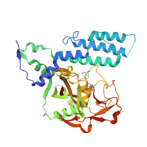
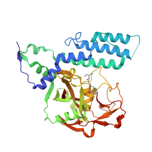
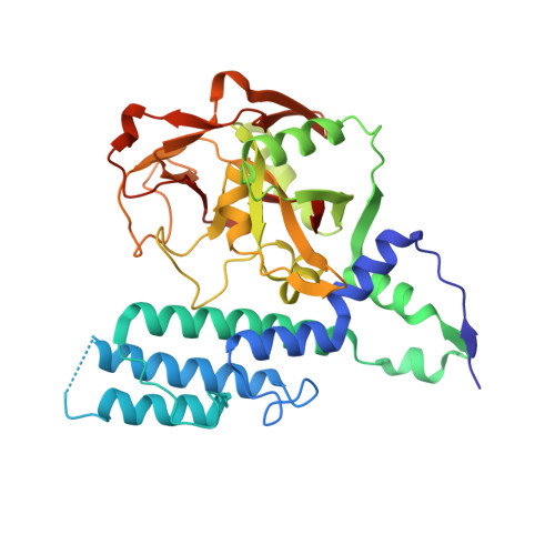
RCSB PDB - 4L7R: Human artd3 (parp3) - catalytic domain in complex with inhibitor ME0400

 4L7R

Human artd3 (parp3) - catalytic domain in complex with inhibitor ME0400


Experimental Data Snapshot

  • Method: X-RAY DIFFRACTION
  • Resolution: 2.20 Å
  • R-Value Free: 0.227 
  • R-Value Work: 0.175 
  • R-Value Observed: 0.179 

Starting Model: experimental
View more details

wwPDB Validation   3D Report Full Report


Ligand Structure Quality Assessment 

Created with Raphaël 2.3.0Worse 01 BetterLigand structure goodness of fit to experimental dataBest fitted M00Click on this verticalbar to view details

This is version 1.1 of the entry. See complete history


Literature

Chemical Probes to Study ADP-Ribosylation: Synthesis and Biochemical Evaluation of Inhibitors of the Human ADP-Ribosyltransferase ARTD3/PARP3.

Lindgren, A.E.Karlberg, T.Ekblad, T.Spjut, S.Thorsell, A.G.Andersson, C.D.Nhan, T.T.Hellsten, V.Weigelt, J.Linusson, A.Schuler, H.Elofsson, M.

(2013) J Med Chem 56: 9556-9568

  • DOI: https://doi.org/10.1021/jm401394u
  • Primary Citation of Related Structures:  
    4L6Z, 4L70, 4L7L, 4L7N, 4L7O, 4L7P, 4L7R, 4L7U

  • PubMed Abstract: 

    The racemic 3-(4-oxo-3,4-dihydroquinazolin-2-yl)-N-[1-(pyridin-2-yl)ethyl]propanamide, 1, has previously been identified as a potent but unselective inhibitor of diphtheria toxin-like ADP-ribosyltransferase 3 (ARTD3). Herein we describe synthesis and evaluation of 55 compounds in this class. It was found that the stereochemistry is of great importance for both selectivity and potency and that substituents on the phenyl ring resulted in poor solubility. Certain variations at the meso position were tolerated and caused a large shift in the binding pose. Changes to the ethylene linker that connects the quinazolinone to the amide were also investigated but proved detrimental to binding. By combination of synthetic organic chemistry and structure-based design, two selective inhibitors of ARTD3 were discovered.


  • Organizational Affiliation

    Department of Chemistry, Umeå University , SE-90187 Umeå, Sweden.


Macromolecules
Find similar proteins by:  (by identity cutoff)  |  3D Structure
Entity ID: 1
MoleculeChains Sequence LengthOrganismDetailsImage
Poly [ADP-ribose] polymerase 3357Homo sapiensMutation(s): 0 
Gene Names: ADPRT3ADPRTL3PARP3
EC: 2.4.2.30 (PDB Primary Data), 2.4.2 (UniProt)
UniProt & NIH Common Fund Data Resources
Find proteins for Q9Y6F1 (Homo sapiens)
Explore Q9Y6F1 
Go to UniProtKB:  Q9Y6F1
PHAROS:  Q9Y6F1
GTEx:  ENSG00000041880 
Entity Groups  
Sequence Clusters30% Identity50% Identity70% Identity90% Identity95% Identity100% Identity
UniProt GroupQ9Y6F1
Sequence Annotations
Expand
  • Reference Sequence
Small Molecules
Ligands 1 Unique
IDChains Name / Formula / InChI Key2D Diagram3D Interactions
M00
Query on M00

Download Ideal Coordinates CCD File 
B [auth A]N-[(2S)-1-hydroxybutan-2-yl]-3-(4-oxo-3,4-dihydroquinazolin-2-yl)propanamide
C15 H19 N3 O3
INPZIMSQNZGLHQ-JTQLQIEISA-N
Binding Affinity Annotations 
IDSourceBinding Affinity
M00 BindingDB:  4L7R IC50: 9600 (nM) from 1 assay(s)
Experimental Data & Validation

Experimental Data

  • Method: X-RAY DIFFRACTION
  • Resolution: 2.20 Å
  • R-Value Free: 0.227 
  • R-Value Work: 0.175 
  • R-Value Observed: 0.179 
  • Space Group: P 1 21 1
Unit Cell:
Length ( Å )Angle ( ˚ )
a = 55.324α = 90
b = 56.456β = 112.97
c = 56.475γ = 90
Software Package:
Software NamePurpose
MxCuBEdata collection
MOLREPphasing
BUSTERrefinement
XDSdata reduction
XSCALEdata scaling

Structure Validation

View Full Validation Report


Ligand Structure Quality Assessment 

Created with Raphaël 2.3.0Worse 01 BetterLigand structure goodness of fit to experimental dataBest fitted M00Click on this verticalbar to view details

Entry History 

Deposition Data

Revision History  (Full details and data files)

  • Version 1.0: 2014-02-19
    Type: Initial release
  • Version 1.1: 2023-09-20
    Changes: Data collection, Database references, Derived calculations, Refinement description