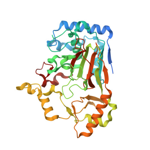
4M2C

Crystal structure of non-heme iron oxygenase OrfP in complex with Fe and D-Arg


Experimental Data Snapshot

  • Method: X-RAY DIFFRACTION
  • Resolution: 2.35 Å
  • R-Value Free: 0.276 
  • R-Value Work: 0.223 
  • R-Value Observed: 0.226 

Starting Model: experimental
View more details

wwPDB Validation   3D Report Full Report


Ligand Structure Quality Assessment 


This is version 1.2 of the entry. See complete history


Literature

Biosynthesis of streptolidine involved two unexpected intermediates produced by a dihydroxylase and a cyclase through unusual mechanisms.

Chang, C.Y.Lyu, S.Y.Liu, Y.C.Hsu, N.S.Wu, C.C.Tang, C.F.Lin, K.H.Ho, J.Y.Wu, C.J.Tsai, M.D.Li, T.L.

(2014) Angew Chem Int Ed Engl 53: 1943-1948

  • DOI: https://doi.org/10.1002/anie.201307989
  • Primary Citation of Related Structures:  
    4M23, 4M25, 4M26, 4M27, 4M2C, 4M2E, 4M2F, 4M2G, 4M2I, 4M2J, 4M2K, 4M2M, 4NE0

  • PubMed Abstract: 

    Streptothricin-F (STT-F), one of the early-discovered antibiotics, consists of three components, a β-lysine homopolymer, an aminosugar D-gulosamine, and an unusual bicyclic streptolidine. The biosynthesis of streptolidine is a long-lasting but unresolved puzzle. Herein, a combination of genetic/biochemical/structural approaches was used to unravel this problem. The STT gene cluster was first sequenced from a Streptomyces variant BCRC 12163, wherein two gene products OrfP and OrfR were characterized in vitro to be a dihydroxylase and a cyclase, respectively. Thirteen high-resolution crystal structures for both enzymes in different reaction intermediate states were snapshotted to help elucidate their catalytic mechanisms. OrfP catalyzes an Fe(II) -dependent double hydroxylation reaction converting L-Arg into (3R,4R)-(OH)2 -L-Arg via (3S)-OH-L-Arg, while OrfR catalyzes an unusual PLP-dependent elimination/addition reaction cyclizing (3R,4R)-(OH)2 -L-Arg to the six-membered (4R)-OH-capreomycidine. The biosynthetic mystery finally comes to light as the latter product was incorporation into STT-F by a feeding experiment.


  • Organizational Affiliation

    Genomics Research Center, Academia Sinica, 128 Academia Road, Section 2, Nankang, Taipei 115 (Taiwan).


Macromolecules
Find similar proteins by:  (by identity cutoff)  |  3D Structure
Entity ID: 1
MoleculeChains Sequence LengthOrganismDetailsImage
L-arginine beta-hydroxylase
A, B, C, D
364Streptomyces lavendulae subsp. lavendulaeMutation(s): 0 
EC: 1.14.11.41
UniProt
Find proteins for G9MBV2 (Streptomyces lavendulae subsp. lavendulae)
Explore G9MBV2 
Go to UniProtKB:  G9MBV2
Entity Groups  
Sequence Clusters30% Identity50% Identity70% Identity90% Identity95% Identity100% Identity
UniProt GroupG9MBV2
Sequence Annotations
Expand
  • Reference Sequence
Experimental Data & Validation

Experimental Data

  • Method: X-RAY DIFFRACTION
  • Resolution: 2.35 Å
  • R-Value Free: 0.276 
  • R-Value Work: 0.223 
  • R-Value Observed: 0.226 
  • Space Group: P 1 21 1
Unit Cell:
Length ( Å )Angle ( ˚ )
a = 68.034α = 90
b = 116.369β = 91.44
c = 95.8γ = 90
Software Package:
Software NamePurpose
HKL-2000data collection
MOLREPphasing
REFMACrefinement
HKL-2000data reduction
HKL-2000data scaling

Structure Validation

View Full Validation Report


Ligand Structure Quality Assessment 


Entry History 

Deposition Data

Revision History  (Full details and data files)

  • Version 1.0: 2014-06-25
    Type: Initial release
  • Version 1.1: 2018-07-18
    Changes: Data collection
  • Version 1.2: 2023-11-08
    Changes: Data collection, Database references, Derived calculations, Refinement description