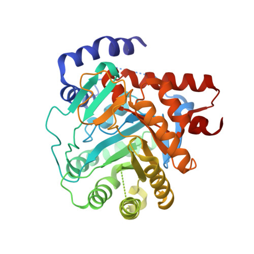
5MUT

Crystal structure of potent human Dihydroorotate Dehydrogenase inhibitors based on hydroxylated azole scaffolds


Experimental Data Snapshot

  • Method: X-RAY DIFFRACTION
  • Resolution: 1.75 Å
  • R-Value Free: 0.198 
  • R-Value Work: 0.168 
  • R-Value Observed: 0.170 

Starting Model: experimental
View more details

wwPDB Validation   3D Report Full Report


Ligand Structure Quality Assessment 


This is version 1.1 of the entry. See complete history


Literature

Design, synthesis, biological evaluation and X-ray structural studies of potent human dihydroorotate dehydrogenase inhibitors based on hydroxylated azole scaffolds.

Sainas, S.Pippione, A.C.Giorgis, M.Lupino, E.Goyal, P.Ramondetti, C.Buccinna, B.Piccinini, M.Braga, R.C.Andrade, C.H.Andersson, M.Moritzer, A.C.Friemann, R.Mensa, S.Al-Kadaraghi, S.Boschi, D.Lolli, M.L.

(2017) Eur J Med Chem 129: 287-302

  • DOI: https://doi.org/10.1016/j.ejmech.2017.02.017
  • Primary Citation of Related Structures:  
    5MUT, 5MVC, 5MVD

  • PubMed Abstract: 

    A new generation of potent hDHODH inhibitors designed by a scaffold-hopping replacement of the quinolinecarboxylate moiety of brequinar, one of the most potent known hDHODH inhibitors, is presented here. Their general structure is characterized by a biphenyl moiety joined through an amide bridge with an acidic hydroxyazole scaffold (hydroxylated thiadiazole, pyrazole and triazole). Molecular modelling suggested that these structures should adopt a brequinar-like binding mode involving interactions with subsites 1, 2 and 4 of the hDHODH binding site. Initially, the inhibitory activity of the compounds was studied on recombinant hDHODH. The most potent compound of the series in the enzymatic assays was the thiadiazole analogue 4 (IC 50 16 nM). The activity was found to be dependent on the fluoro substitution pattern at the biphenyl moiety as well as on the choice/substitution of the heterocyclic ring. Structure determination of hDHODH co-crystallized with one representative compound from each series (4, 5 and 6) confirmed the brequinar-like binding mode as suggested by modelling. The specificity of the observed effects of the compound series was tested in cell-based assays for antiproliferation activity using Jurkat cells and PHA-stimulated PBMC. These tests were also verified by addition of exogenous uridine to the culture medium. In particular, the triazole analogue 6 (IC 50 against hDHODH: 45 nM) exerted potent in vitro antiproliferative and immunosuppressive activity without affecting cell survival.


  • Organizational Affiliation

    Department of Science and Drug Technology, University of Torino, via Pietro Giuria 9, 10125 Torino, Italy.


Macromolecules
Find similar proteins by:  (by identity cutoff)  |  3D Structure
Entity ID: 1
MoleculeChains Sequence LengthOrganismDetailsImage
Dihydroorotate dehydrogenase (quinone), mitochondrial365Homo sapiensMutation(s): 0 
Gene Names: DHODH
EC: 1.3.5.2
UniProt & NIH Common Fund Data Resources
Find proteins for Q02127 (Homo sapiens)
Explore Q02127 
Go to UniProtKB:  Q02127
PHAROS:  Q02127
GTEx:  ENSG00000102967 
Entity Groups  
Sequence Clusters30% Identity50% Identity70% Identity90% Identity95% Identity100% Identity
UniProt GroupQ02127
Sequence Annotations
Expand
  • Reference Sequence
Small Molecules
Ligands 5 Unique
IDChains Name / Formula / InChI Key2D Diagram3D Interactions
FMN
Query on FMN

Download Ideal Coordinates CCD File 
B [auth A]FLAVIN MONONUCLEOTIDE
C17 H21 N4 O9 P
FVTCRASFADXXNN-SCRDCRAPSA-N
HYT
Query on HYT

Download Ideal Coordinates CCD File 
D [auth A]2-methyl-5-oxidanyl-~{N}-[2,3,5,6-tetrakis(fluoranyl)-4-phenyl-phenyl]-1,2,3-triazole-4-carboxamide
C16 H10 F4 N4 O2
NRGHNOMCGATBLG-UHFFFAOYSA-N
ORO
Query on ORO

Download Ideal Coordinates CCD File 
C [auth A]OROTIC ACID
C5 H4 N2 O4
PXQPEWDEAKTCGB-UHFFFAOYSA-N
ACT
Query on ACT

Download Ideal Coordinates CCD File 
E [auth A],
F [auth A]
ACETATE ION
C2 H3 O2
QTBSBXVTEAMEQO-UHFFFAOYSA-M
CL
Query on CL

Download Ideal Coordinates CCD File 
G [auth A],
H [auth A],
I [auth A]
CHLORIDE ION
Cl
VEXZGXHMUGYJMC-UHFFFAOYSA-M
Binding Affinity Annotations 
IDSourceBinding Affinity
HYT BindingDB:  5MUT IC50: 45 (nM) from 1 assay(s)
Experimental Data & Validation

Experimental Data

  • Method: X-RAY DIFFRACTION
  • Resolution: 1.75 Å
  • R-Value Free: 0.198 
  • R-Value Work: 0.168 
  • R-Value Observed: 0.170 
  • Space Group: P 32 2 1
Unit Cell:
Length ( Å )Angle ( ˚ )
a = 91.08α = 90
b = 91.08β = 90
c = 122.522γ = 120
Software Package:
Software NamePurpose
MOSFLMdata reduction
PHASERphasing
PHENIXrefinement
PDB_EXTRACTdata extraction
SCALAdata scaling

Structure Validation

View Full Validation Report


Ligand Structure Quality Assessment 


Entry History 

Deposition Data

Revision History  (Full details and data files)

  • Version 1.0: 2017-03-08
    Type: Initial release
  • Version 1.1: 2024-01-17
    Changes: Data collection, Database references, Refinement description