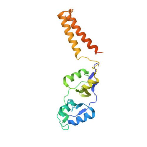
5NPL

Crystal structure of hexameric CBS-CP12 protein from bloom-forming cyanobacteria, Yb-derivative at 2.8 A resolution


Experimental Data Snapshot

  • Method: X-RAY DIFFRACTION
  • Resolution: 2.79 Å
  • R-Value Free: 0.219 
  • R-Value Work: 0.186 
  • R-Value Observed: 0.188 

wwPDB Validation   3D Report Full Report


Ligand Structure Quality Assessment 


This is version 1.3 of the entry. See complete history


Literature

Structural and functional insights into the unique CBS-CP12 fusion protein family in cyanobacteria.

Hackenberg, C.Hakanpaa, J.Cai, F.Antonyuk, S.Eigner, C.Meissner, S.Laitaoja, M.Janis, J.Kerfeld, C.A.Dittmann, E.Lamzin, V.S.

(2018) Proc Natl Acad Sci U S A 115: 7141-7146

  • DOI: https://doi.org/10.1073/pnas.1806668115
  • Primary Citation of Related Structures:  
    5NMU, 5NPL, 5NVD

  • PubMed Abstract: 

    Cyanobacteria are important photosynthetic organisms inhabiting a range of dynamic environments. This phylum is distinctive among photosynthetic organisms in containing genes encoding uncharacterized cystathionine β-synthase (CBS)-chloroplast protein (CP12) fusion proteins. These consist of two domains, each recognized as stand-alone photosynthetic regulators with different functions described in cyanobacteria (CP12) and plants (CP12 and CBSX). Here we show that CBS-CP12 fusion proteins are encoded in distinct gene neighborhoods, several unrelated to photosynthesis. Most frequently, CBS-CP12 genes are in a gene cluster with thioredoxin A (TrxA), which is prevalent in bloom-forming, marine symbiotic, and benthic mat cyanobacteria. Focusing on a CBS-CP12 from Microcystis aeruginosa PCC 7806 encoded in a gene cluster with TrxA, we reveal that the domain fusion led to the formation of a hexameric protein. We show that the CP12 domain is essential for hexamerization and contains an ordered, previously structurally uncharacterized N-terminal region. We provide evidence that CBS-CP12, while combining properties of both regulatory domains, behaves different from CP12 and plant CBSX. It does not form a ternary complex with phosphoribulokinase (PRK) and glyceraldehyde-3-phosphate dehydrogenase. Instead, CBS-CP12 decreases the activity of PRK in an AMP-dependent manner. We propose that the novel domain architecture and oligomeric state of CBS-CP12 expand its regulatory function beyond those of CP12 in cyanobacteria.


  • Organizational Affiliation

    European Molecular Biology Laboratory (EMBL), Deutsches Elektronen-Synchrotron (DESY), 22607 Hamburg, Germany; [email protected] [email protected].


Macromolecules
Find similar proteins by:  (by identity cutoff)  |  3D Structure
Entity ID: 1
MoleculeChains Sequence LengthOrganismDetailsImage
Similar to tr|Q8YYT1|Q8YYT1
A, B, C
208Microcystis aeruginosa PCC 7806Mutation(s): 0 
Gene Names: IPF_2164
UniProt
Find proteins for A8YJ50 (Microcystis aeruginosa (strain PCC 7806))
Explore A8YJ50 
Go to UniProtKB:  A8YJ50
Entity Groups  
Sequence Clusters30% Identity50% Identity70% Identity90% Identity95% Identity100% Identity
UniProt GroupA8YJ50
Sequence Annotations
Expand
  • Reference Sequence
Small Molecules
Ligands 2 Unique
IDChains Name / Formula / InChI Key2D Diagram3D Interactions
DO3
Query on DO3

Download Ideal Coordinates CCD File 
E [auth A],
G [auth A]
10-((2R)-2-HYDROXYPROPYL)-1,4,7,10-TETRAAZACYCLODODECANE 1,4,7-TRIACETIC ACID
C17 H32 N4 O7
IQUHNCOJRJBMSU-CQSZACIVSA-N
YB
Query on YB

Download Ideal Coordinates CCD File 
D [auth A]
F [auth A]
H [auth A]
I [auth A]
J [auth A]
D [auth A],
F [auth A],
H [auth A],
I [auth A],
J [auth A],
K [auth A],
L [auth B],
M [auth B],
N [auth B],
O [auth C],
P [auth C],
Q [auth C],
R [auth C],
S [auth C],
T [auth C],
U [auth C],
V [auth C]
YTTERBIUM (III) ION
Yb
AWSFICBXMUKWSK-UHFFFAOYSA-N
Experimental Data & Validation

Experimental Data

  • Method: X-RAY DIFFRACTION
  • Resolution: 2.79 Å
  • R-Value Free: 0.219 
  • R-Value Work: 0.186 
  • R-Value Observed: 0.188 
  • Space Group: C 2 2 21
Unit Cell:
Length ( Å )Angle ( ˚ )
a = 79.89α = 90
b = 120.734β = 90
c = 117.548γ = 90
Software Package:
Software NamePurpose
REFMACrefinement
XDSdata reduction
XDSdata scaling
CRANK2phasing

Structure Validation

View Full Validation Report


Ligand Structure Quality Assessment 


Entry History 

Deposition Data

Revision History  (Full details and data files)

  • Version 1.0: 2018-05-30
    Type: Initial release
  • Version 1.1: 2018-06-20
    Changes: Data collection, Database references
  • Version 1.2: 2018-07-11
    Changes: Data collection, Database references
  • Version 1.3: 2024-11-06
    Changes: Data collection, Database references, Refinement description, Structure summary