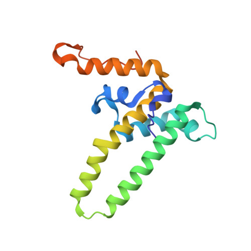
5WRE

Hepatitis B virus core protein Y132A mutant in complex with heteroaryldihydropyrimidine (HAP_R01)


Experimental Data Snapshot

  • Method: X-RAY DIFFRACTION
  • Resolution: 1.95 Å
  • R-Value Free: 0.239 
  • R-Value Work: 0.207 
  • R-Value Observed: 0.208 

Starting Model: experimental
View more details

wwPDB Validation   3D Report Full Report


Ligand Structure Quality Assessment 


This is version 1.3 of the entry. See complete history


Literature

Heteroaryldihydropyrimidine (HAP) and Sulfamoylbenzamide (SBA) Inhibit Hepatitis B Virus Replication by Different Molecular Mechanisms.

Zhou, Z.Hu, T.Zhou, X.Wildum, S.Garcia-Alcalde, F.Xu, Z.Wu, D.Mao, Y.Tian, X.Zhou, Y.Shen, F.Zhang, Z.Tang, G.Najera, I.Yang, G.Shen, H.C.Young, J.A.Qin, N.

(2017) Sci Rep 7: 42374-42374

  • DOI: https://doi.org/10.1038/srep42374
  • Primary Citation of Related Structures:  
    5T2P, 5WRE, 5WTW

  • PubMed Abstract: 

    Heteroaryldihydropyrimidine (HAP) and sulfamoylbenzamide (SBA) are promising non-nucleos(t)ide HBV replication inhibitors. HAPs are known to promote core protein mis-assembly, but the molecular mechanism of abnormal assembly is still elusive. Likewise, the assembly status of core protein induced by SBA remains unknown. Here we show that SBA, unlike HAP, does not promote core protein mis-assembly. Interestingly, two reference compounds HAP_R01 and SBA_R01 bind to the same pocket at the dimer-dimer interface in the crystal structures of core protein Y132A hexamer. The striking difference lies in a unique hydrophobic subpocket that is occupied by the thiazole group of HAP_R01, but is unperturbed by SBA_R01. Photoaffinity labeling confirms the HAP_R01 binding pose at the dimer-dimer interface on capsid and suggests a new mechanism of HAP-induced mis-assembly. Based on the common features in crystal structures we predict that T33 mutations generate similar susceptibility changes to both compounds. In contrast, mutations at positions in close contact with HAP-specific groups (P25A, P25S, or V124F) only reduce susceptibility to HAP_R01, but not to SBA_R01. Thus, HAP and SBA are likely to have distinctive resistance profiles. Notably, P25S and V124F substitutions exist in low-abundance quasispecies in treatment-naïve patients, suggesting potential clinical relevance.


  • Organizational Affiliation

    Roche Pharma Research and Early Development, Chemical Biology, Roche Innovation Center Shanghai, Shanghai, 201203, China.


Macromolecules
Find similar proteins by:  (by identity cutoff)  |  3D Structure
Entity ID: 1
MoleculeChains Sequence LengthOrganismDetailsImage
Core protein
A, B, C, D, E
A, B, C, D, E, F
155Hepatitis B virusMutation(s): 1 
UniProt
Find proteins for L7R9I1 (Hepatitis B virus)
Explore L7R9I1 
Go to UniProtKB:  L7R9I1
Entity Groups  
Sequence Clusters30% Identity50% Identity70% Identity90% Identity95% Identity100% Identity
UniProt GroupL7R9I1
Sequence Annotations
Expand
  • Reference Sequence
Small Molecules
Ligands 4 Unique
IDChains Name / Formula / InChI Key2D Diagram3D Interactions
7TL
Query on 7TL

Download Ideal Coordinates CCD File 
G [auth A]
K [auth B]
P [auth C]
S [auth D]
V [auth E]
G [auth A],
K [auth B],
P [auth C],
S [auth D],
V [auth E],
Z [auth F]
(2S)-1-[[(4R)-4-(2-chloranyl-4-fluoranyl-phenyl)-5-methoxycarbonyl-2-(1,3-thiazol-2-yl)-1,4-dihydropyrimidin-6-yl]methyl]-4,4-bis(fluoranyl)pyrrolidine-2-carboxylic acid
C21 H18 Cl F3 N4 O4 S
PEWCUEKNYZSVSW-HOCLYGCPSA-N
GOL
Query on GOL

Download Ideal Coordinates CCD File 
AA [auth F]
BA [auth F]
N [auth B]
O [auth B]
R [auth C]
AA [auth F],
BA [auth F],
N [auth B],
O [auth B],
R [auth C],
Y [auth E]
GLYCEROL
C3 H8 O3
PEDCQBHIVMGVHV-UHFFFAOYSA-N
IPA
Query on IPA

Download Ideal Coordinates CCD File 
H [auth A]
I [auth A]
J [auth A]
Q [auth C]
T [auth D]
H [auth A],
I [auth A],
J [auth A],
Q [auth C],
T [auth D],
W [auth E],
X [auth E]
ISOPROPYL ALCOHOL
C3 H8 O
KFZMGEQAYNKOFK-UHFFFAOYSA-N
CL
Query on CL

Download Ideal Coordinates CCD File 
L [auth B],
M [auth B],
U [auth D]
CHLORIDE ION
Cl
VEXZGXHMUGYJMC-UHFFFAOYSA-M
Binding Affinity Annotations 
IDSourceBinding Affinity
7TL BindingDB:  5WRE EC50: 3 (nM) from 1 assay(s)
Experimental Data & Validation

Experimental Data

  • Method: X-RAY DIFFRACTION
  • Resolution: 1.95 Å
  • R-Value Free: 0.239 
  • R-Value Work: 0.207 
  • R-Value Observed: 0.208 
  • Space Group: P 1
Unit Cell:
Length ( Å )Angle ( ˚ )
a = 65.241α = 68.28
b = 66.93β = 69.02
c = 87.071γ = 83.62
Software Package:
Software NamePurpose
DENZOdata collection
SCALEPACKdata scaling
PHENIXrefinement
PDB_EXTRACTdata extraction
HKL-2000data reduction
PHASERphasing

Structure Validation

View Full Validation Report


Ligand Structure Quality Assessment 


Entry History 

Deposition Data

Revision History  (Full details and data files)

  • Version 1.0: 2017-02-22
    Type: Initial release
  • Version 1.1: 2020-01-01
    Changes: Database references
  • Version 1.2: 2023-11-08
    Changes: Data collection, Database references, Refinement description
  • Version 1.3: 2024-10-23
    Changes: Structure summary