5ZJ6

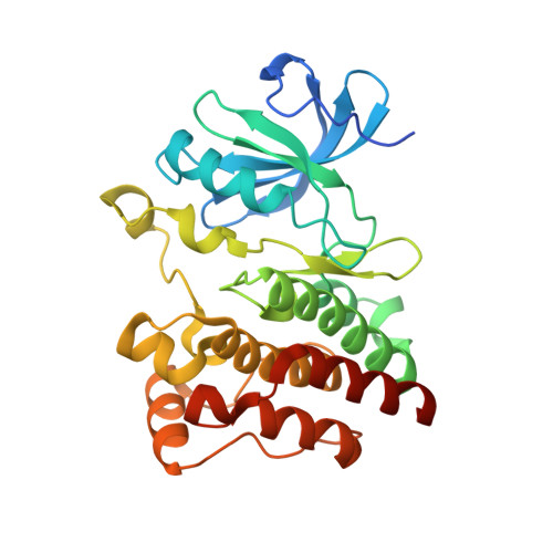
Crystal structure of HCK kinase complexed with a pyrrolo-pyrimidine inhibitor 7-[trans-4-(4-methylpiperazin-1-yl)cyclohexyl]-5-(4-phenoxyphenyl)-7H-pyrrolo[2,3-d]pyrimidin-4-amine


Experimental Data Snapshot

  • Method: X-RAY DIFFRACTION
  • Resolution: 1.70 Å
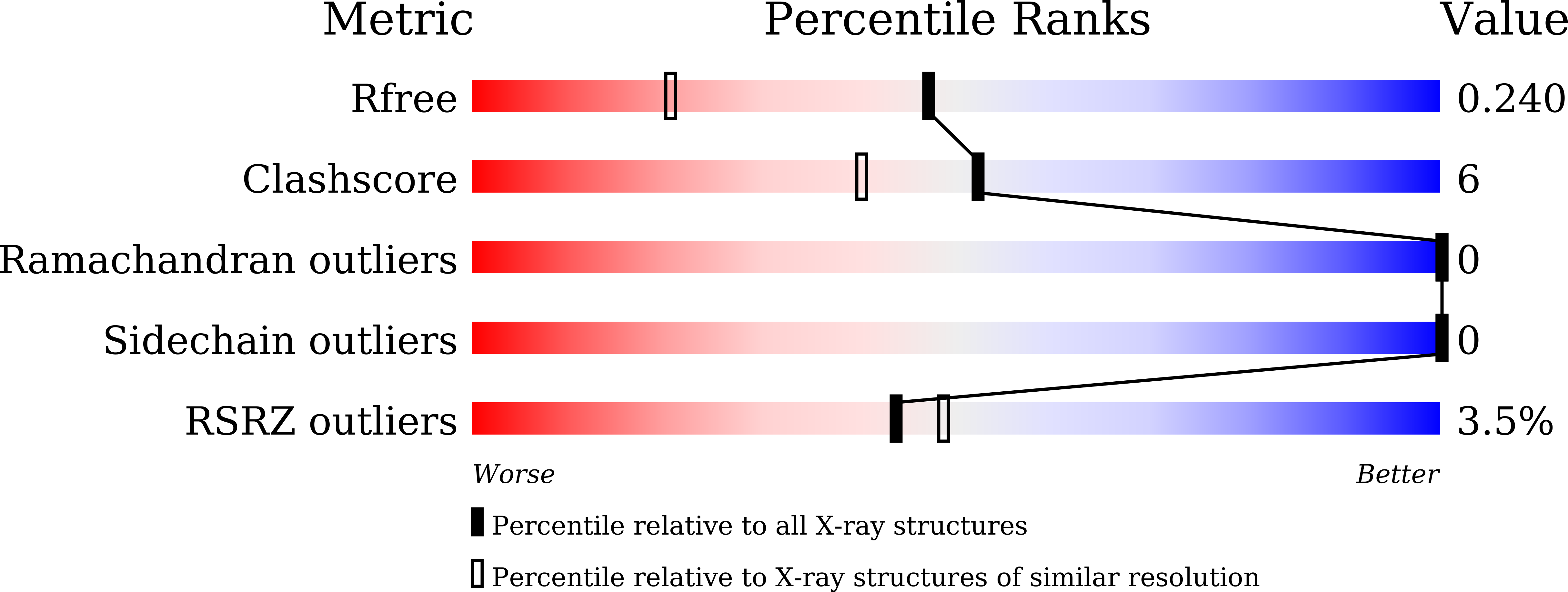
  • R-Value Free: 0.240 
  • R-Value Work: 0.207 
  • R-Value Observed: 0.208 

Starting Model: experimental
View more details

wwPDB Validation   3D Report Full Report


Ligand Structure Quality Assessment 


This is version 1.2 of the entry. See complete history


Literature

Phosphorylated and non-phosphorylated HCK kinase domains produced by cell-free protein expression.

Katsura, K.Tomabechi, Y.Matsuda, T.Yonemochi, M.Mikuni, J.Ohsawa, N.Terada, T.Yokoyama, S.Kukimoto-Niino, M.Takemoto, C.Shirouzu, M.

(2018) Protein Expr Purif 150: 92-99

  • DOI: https://doi.org/10.1016/j.pep.2018.05.005
  • Primary Citation of Related Structures:  
    5ZJ6

  • PubMed Abstract: 

    Since phosphorylation is involved in various physiological events, kinases and interacting factors can be potential targets for drug discovery. For the development and improvement of inhibitors from the point of view of mechanistic enzymology, a cell-free protein synthesis system would be advantageous, since it could prepare mutant proteins easily. However, especially in the case of protein kinase, product solubility remains one of the major challenges. To overcome this problem, we prepared a chaperone-supplemented extract from Escherichia coli BL21 cells harboring a plasmid encoding a set of chaperone genes, dnaK, dnaJ, and grpE. We explored cell-disruption procedures and constructed an efficient protein synthesis system. Employing this system, we produced the kinase domain of human hematopoietic cell kinase (HCK) to obtain further structural information about its molecular interaction with one of its inhibitors, previously developed by our group (RK-20449). Lower reaction temperature improved the solubility, and addition of a protein phosphatase (YpoH) facilitated the homogeneous production of the non-phosphorylated kinase domain. Crystals of the purified product were obtained and the kinase-inhibitor complex structure was solved at 1.7 Å resolution. In addition, results of kinase activity measurement, using a synthetic substrate, showed that the kinase activity was facilitated by autophosphorylation at Tyr416, as confirmed by the peptide mass mapping.


  • Organizational Affiliation

    Protein Functional and Structural Biology Team, RIKEN Center for Life Science Technology, 1-7-22 Suehiro-cho, Tsurumi-ku, Yokohama 230-0045, Japan.


Macromolecules
Find similar proteins by:  (by identity cutoff)  |  3D Structure
Entity ID: 1
MoleculeChains Sequence LengthOrganismDetailsImage
Tyrosine-protein kinase HCK
A, B
287Homo sapiensMutation(s): 0 
Gene Names: HCK
EC: 2.7.10.2
UniProt & NIH Common Fund Data Resources
Find proteins for P08631 (Homo sapiens)
Explore P08631 
Go to UniProtKB:  P08631
PHAROS:  P08631
GTEx:  ENSG00000101336 
Entity Groups  
Sequence Clusters30% Identity50% Identity70% Identity90% Identity95% Identity100% Identity
UniProt GroupP08631
Sequence Annotations
Expand
  • Reference Sequence
Small Molecules
Ligands 1 Unique
IDChains Name / Formula / InChI Key2D Diagram3D Interactions
VSE
Query on VSE

Download Ideal Coordinates CCD File 
C [auth A],
D [auth B]
7-[trans-4-(4-methylpiperazin-1-yl)cyclohexyl]-5-(4-phenoxyphenyl)-7H-pyrrolo[2,3-d]pyrimidin-4-amine
C29 H34 N6 O
FDVSOQRNTAPCHB-YHBQERECSA-N
Binding Affinity Annotations 
IDSourceBinding Affinity
VSE BindingDB:  5ZJ6 IC50: min: 0.4, max: 3.3 (nM) from 4 assay(s)
Experimental Data & Validation

Experimental Data

  • Method: X-RAY DIFFRACTION
  • Resolution: 1.70 Å
  • R-Value Free: 0.240 
  • R-Value Work: 0.207 
  • R-Value Observed: 0.208 
  • Space Group: P 1
Unit Cell:
Length ( Å )Angle ( ˚ )
a = 42.81α = 87.59
b = 48.02β = 83.09
c = 78.01γ = 63.59
Software Package:
Software NamePurpose
XDSdata reduction
XSCALEdata scaling
PHASERphasing
PHENIXrefinement
PDB_EXTRACTdata extraction

Structure Validation

View Full Validation Report


Ligand Structure Quality Assessment 


Entry History 

Deposition Data

Revision History  (Full details and data files)

  • Version 1.0: 2018-06-06
    Type: Initial release
  • Version 1.1: 2018-06-13
    Changes: Data collection, Database references
  • Version 1.2: 2023-11-22
    Changes: Data collection, Database references, Refinement description