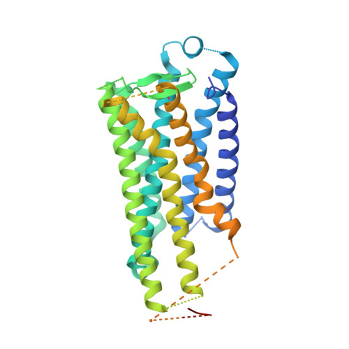
6AK3

Crystal structure of the human prostaglandin E receptor EP3 bound to prostaglandin E2


Experimental Data Snapshot

  • Method: X-RAY DIFFRACTION
  • Resolution: 2.90 Å
  • R-Value Free: 0.295 
  • R-Value Work: 0.251 
  • R-Value Observed: 0.253 

wwPDB Validation   3D Report Full Report


Ligand Structure Quality Assessment 


This is version 1.5 of the entry. See complete history


Literature

Crystal structure of the endogenous agonist-bound prostanoid receptor EP3.

Morimoto, K.Suno, R.Hotta, Y.Yamashita, K.Hirata, K.Yamamoto, M.Narumiya, S.Iwata, S.Kobayashi, T.

(2019) Nat Chem Biol 15: 8-10

  • DOI: https://doi.org/10.1038/s41589-018-0171-8
  • Primary Citation of Related Structures:  
    6AK3

  • PubMed Abstract: 

    Prostanoids are a series of bioactive lipid metabolites that function in an autacoid manner via activation of cognate G-protein-coupled receptors (GPCRs). Here, we report the crystal structure of human prostaglandin (PG) E receptor subtype EP3 bound to endogenous ligand PGE 2 at 2.90 Å resolution. The structure reveals important insights into the activation mechanism of prostanoid receptors and provides a molecular basis for the binding modes of endogenous ligands.


  • Organizational Affiliation

    Department of Cell Biology, Graduate School of Medicine, Kyoto University, Konoe-cho, Yoshida, Sakyo-ku, Kyoto, Japan. [email protected].


Macromolecules
Find similar proteins by:  (by identity cutoff)  |  3D Structure
Entity ID: 1
MoleculeChains Sequence LengthOrganismDetailsImage
Prostaglandin E2 receptor EP3 subtype,Soluble cytochrome b562A [auth B],
B [auth A]
418Homo sapiensEscherichia coliMutation(s): 9 
Gene Names: PTGER3cybC
Membrane Entity: Yes 
UniProt & NIH Common Fund Data Resources
Find proteins for P0ABE7 (Escherichia coli)
Explore P0ABE7 
Go to UniProtKB:  P0ABE7
Find proteins for P43115 (Homo sapiens)
Explore P43115 
Go to UniProtKB:  P43115
PHAROS:  P43115
GTEx:  ENSG00000050628 
Entity Groups  
Sequence Clusters30% Identity50% Identity70% Identity90% Identity95% Identity100% Identity
UniProt GroupsP0ABE7P43115
Sequence Annotations
Expand
  • Reference Sequence
Small Molecules
Binding Affinity Annotations 
IDSourceBinding Affinity
P2E BindingDB:  6AK3 Ki: min: 0.33, max: 5 (nM) from 3 assay(s)
IC50: min: 0.94, max: 3 (nM) from 2 assay(s)
EC50: 2.5 (nM) from 1 assay(s)
Experimental Data & Validation

Experimental Data

Unit Cell:
Length ( Å )Angle ( ˚ )
a = 66.09α = 90
b = 42.28β = 96.09
c = 161.3γ = 90
Software Package:
Software NamePurpose
PHENIXrefinement
XDSdata reduction
XDSdata scaling
PHASERphasing

Structure Validation

View Full Validation Report


Ligand Structure Quality Assessment 


Entry History & Funding Information

Deposition Data


Funding OrganizationLocationGrant Number
Japan Agency for Medical Research and Development (AMED)JapanJP18gm0910007
Japan Agency for Medical Research and Development (AMED)JapanJP18am0101079

Revision History  (Full details and data files)

  • Version 1.0: 2018-12-05
    Type: Initial release
  • Version 1.1: 2018-12-12
    Changes: Data collection, Structure summary
  • Version 1.2: 2018-12-19
    Changes: Data collection, Database references
  • Version 1.3: 2019-01-30
    Changes: Data collection, Database references
  • Version 1.4: 2023-09-06
    Changes: Data collection, Database references
  • Version 1.5: 2024-11-06
    Changes: Structure summary