7XQK

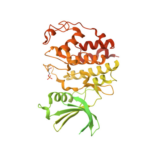
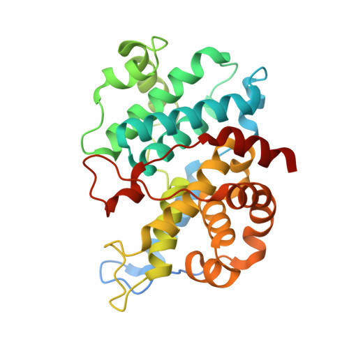
The Crystal Structure of CDK3 and CyclinE1 Complex from Biortus.


Experimental Data Snapshot

  • Method: X-RAY DIFFRACTION
  • Resolution: 2.25 Å
  • R-Value Free: 0.202 
  • R-Value Work: 0.160 

Starting Model: experimental
View more details

wwPDB Validation   3D Report Full Report


This is version 1.3 of the entry. See complete history


Literature

Structural basis of CDK3 activation by cyclin E1 and inhibition by dinaciclib.

Gui, W.Hang, Y.Cheng, W.Gao, M.Wu, J.Ouyang, Z.

(2023) Biochem Biophys Res Commun 662: 126-134

  • DOI: https://doi.org/10.1016/j.bbrc.2023.04.026
  • Primary Citation of Related Structures:  
    7XQK, 8H4R

  • PubMed Abstract: 

    Cell cycle transitions are controlled by multiple cell cycle regulators, especially CDKs. Several CDKs, including CDK1-4 and CDK6, promote cell cycle progression directly. Among them, CDK3 is critically important because it triggers the transitions of G0 to G1 and G1 to S phase through binding to cyclin C and cyclin E1, respectively. In contrast to its highly related homologs, the molecular basis of CDK3 activation remains elusive due to the lack of structural information of CDK3, particularly in cyclin bound form. Here we report the crystal structure of CDK3 in complex with cyclin E1 at 2.25 Å resolution. CDK3 resembles CDK2 in that both adopt a similar fold and bind cyclin E1 in a similar way. The structural discrepancy between CDK3 and CDK2 may reflect their substrate specificity. Profiling a panel of CDK inhibitors reveals that dinaciclib inhibits CDK3-cyclin E1 potently and specifically. The structure of CDK3-cyclin E1 bound to dinaciclib reveals the inhibitory mechanism. The structural and biochemical results uncover the mechanism of CDK3 activation by cyclin E1 and lays a foundation for structural-based drug design.


  • Organizational Affiliation

    Wuxi Biortus Biosciences Co. Ltd, 6 Dongsheng Western Road, Jiangyin, Jiangsu, 214437, China.


Macromolecules
Find similar proteins by:  (by identity cutoff)  |  3D Structure
Entity ID: 1
MoleculeChains Sequence LengthOrganismDetailsImage
Glutathione S-transferase class-mu 26 kDa isozyme,Cyclin-dependent kinase 3533Schistosoma japonicumHomo sapiens
This entity is chimeric
Mutation(s): 0 
EC: 2.5.1.18 (PDB Primary Data), 2.7.11.22 (PDB Primary Data)
UniProt & NIH Common Fund Data Resources
Find proteins for P08515 (Schistosoma japonicum)
Explore P08515 
Go to UniProtKB:  P08515
Find proteins for Q00526 (Homo sapiens)
Explore Q00526 
Go to UniProtKB:  Q00526
PHAROS:  Q00526
GTEx:  ENSG00000250506 
Entity Groups  
Sequence Clusters30% Identity50% Identity70% Identity90% Identity95% Identity100% Identity
UniProt GroupsP08515Q00526
Sequence Annotations
Expand
  • Reference Sequence
Find similar proteins by:  (by identity cutoff)  |  3D Structure
Entity ID: 2
MoleculeChains Sequence LengthOrganismDetailsImage
G1/S-specific cyclin-E1305Homo sapiensMutation(s): 0 
Gene Names: CCNE1CCNE
UniProt & NIH Common Fund Data Resources
Find proteins for P24864 (Homo sapiens)
Explore P24864 
Go to UniProtKB:  P24864
PHAROS:  P24864
GTEx:  ENSG00000105173 
Entity Groups  
Sequence Clusters30% Identity50% Identity70% Identity90% Identity95% Identity100% Identity
UniProt GroupP24864
Sequence Annotations
Expand
  • Reference Sequence
Small Molecules
Ligands 3 Unique
IDChains Name / Formula / InChI Key2D Diagram3D Interactions
MES
Query on MES

Download Ideal Coordinates CCD File 
O [auth B]2-(N-MORPHOLINO)-ETHANESULFONIC ACID
C6 H13 N O4 S
SXGZJKUKBWWHRA-UHFFFAOYSA-N
SO4
Query on SO4

Download Ideal Coordinates CCD File 
C [auth A]
E [auth B]
F [auth B]
G [auth B]
H [auth B]
C [auth A],
E [auth B],
F [auth B],
G [auth B],
H [auth B],
I [auth B],
J [auth B],
K [auth B],
L [auth B],
M [auth B],
N [auth B]
SULFATE ION
O4 S
QAOWNCQODCNURD-UHFFFAOYSA-L
GOL
Query on GOL

Download Ideal Coordinates CCD File 
D [auth A],
P [auth B],
Q [auth B]
GLYCEROL
C3 H8 O3
PEDCQBHIVMGVHV-UHFFFAOYSA-N
Modified Residues  1 Unique
IDChains TypeFormula2D DiagramParent
TPO
Query on TPO
A
L-PEPTIDE LINKINGC4 H10 N O6 PTHR
Experimental Data & Validation

Experimental Data

  • Method: X-RAY DIFFRACTION
  • Resolution: 2.25 Å
  • R-Value Free: 0.202 
  • R-Value Work: 0.160 
  • Space Group: P 63
Unit Cell:
Length ( Å )Angle ( ˚ )
a = 154.154α = 90
b = 154.154β = 90
c = 76.675γ = 120
Software Package:
Software NamePurpose
XDSdata reduction
Aimlessdata scaling
PHASERphasing
REFMACrefinement

Structure Validation

View Full Validation Report


Entry History & Funding Information

Deposition Data


Funding OrganizationLocationGrant Number
Not funded--

Revision History  (Full details and data files)

  • Version 1.0: 2023-05-17
    Type: Initial release
  • Version 1.1: 2023-11-29
    Changes: Data collection, Refinement description
  • Version 1.2: 2024-11-13
    Changes: Structure summary
  • Version 1.3: 2024-11-27
    Changes: Database references