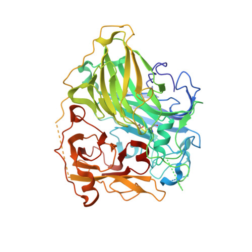
7Y8C

Crystal structure of CotA laccase complexed with syringaldehyde


Experimental Data Snapshot

  • Method: X-RAY DIFFRACTION
  • Resolution: 2.00 Å
  • R-Value Free: 0.219 
  • R-Value Work: 0.172 
  • R-Value Observed: 0.174 

Starting Model: experimental
View more details

wwPDB Validation   3D Report Full Report


Ligand Structure Quality Assessment 


This is version 1.4 of the entry. See complete history


Literature

Molecular insights into substrate promiscuity of CotA laccase catalyzing lignin-phenol derivatives.

Li, J.Liu, Z.Zhao, J.Wang, G.Xie, T.

(2023) Int J Biol Macromol 256: 128487-128487

  • DOI: https://doi.org/10.1016/j.ijbiomac.2023.128487
  • Primary Citation of Related Structures:  
    7Y8B, 7Y8C

  • PubMed Abstract: 

    CotA laccases are multicopper oxidases known for promiscuously oxidizing a broad range of substrates. However, studying substrate promiscuity is limited by the complexity of electron transfer (ET) between substrates and laccases. Here, a systematic analysis of factors affecting ET including electron donor acceptor coupling (Η DA ), driving force (ΔG) and reorganization energy (λ) was done. Catalysis rates of syringic acid (SA), syringaldehyde (SAD) and acetosyringone (AS) (kcat(SAD) > kcat(SA) > kcat(AS)) are not entirely dependent on the ability to form phenol radicals indicated by ΔG and λ calculated by Density Functional Theory (SA < SAD ≈ AS). In determined CotA/SA and CotA/SAD structures, SA and SAD bound at 3.9 and 3.7 Å away from T1 Cu coordinating His419 ensuring a similar Η DA . Abilities of substrate to form phenol radicals could mainly account for difference between kcat(SAD) and kcat(SA). Furthermore, substrate pocket is solvent exposed at the para site of substrate's phenol hydroxyl, which would destabilize binding of AS in the same orientation and position resulting in low kcat. Our results indicated shallow partially covered binding site with propensity of amino acids distribution might help CotA discriminate lignin-phenol derivatives. These findings give new insights for developing specific catalysts for industrial application.


  • Organizational Affiliation

    Key Laboratory of Environmental and Applied Microbiology, Chengdu Institute of Biology, Chinese Academy of Sciences, Chengdu 610041, China; Key Laboratory of Environmental Microbiology of Sichuan Province, Chengdu 610041, China; University of Chinese Academy of Sciences, Beijing 100049, China.


Macromolecules
Find similar proteins by:  (by identity cutoff)  |  3D Structure
Entity ID: 1
MoleculeChains Sequence LengthOrganismDetailsImage
Spore coat protein A
A, B
513Bacillus subtilis subsp. subtilis str. 168Mutation(s): 0 
Gene Names: cotApigBSU06300
EC: 1.10.3.2 (UniProt), 1.3.3.5 (UniProt)
UniProt
Find proteins for P07788 (Bacillus subtilis (strain 168))
Explore P07788 
Go to UniProtKB:  P07788
Entity Groups  
Sequence Clusters30% Identity50% Identity70% Identity90% Identity95% Identity100% Identity
UniProt GroupP07788
Sequence Annotations
Expand
  • Reference Sequence
Small Molecules
Ligands 3 Unique
IDChains Name / Formula / InChI Key2D Diagram3D Interactions
IJV (Subject of Investigation/LOI)
Query on IJV

Download Ideal Coordinates CCD File 
L [auth B]3,5-dimethoxy-4-oxidanyl-benzaldehyde
C9 H10 O4
KCDXJAYRVLXPFO-UHFFFAOYSA-N
PGE
Query on PGE

Download Ideal Coordinates CCD File 
K [auth B]TRIETHYLENE GLYCOL
C6 H14 O4
ZIBGPFATKBEMQZ-UHFFFAOYSA-N
CU
Query on CU

Download Ideal Coordinates CCD File 
C [auth A]
D [auth A]
E [auth A]
F [auth A]
G [auth B]
C [auth A],
D [auth A],
E [auth A],
F [auth A],
G [auth B],
H [auth B],
I [auth B],
J [auth B]
COPPER (II) ION
Cu
JPVYNHNXODAKFH-UHFFFAOYSA-N
Experimental Data & Validation

Experimental Data

  • Method: X-RAY DIFFRACTION
  • Resolution: 2.00 Å
  • R-Value Free: 0.219 
  • R-Value Work: 0.172 
  • R-Value Observed: 0.174 
  • Space Group: P 1 21 1
Unit Cell:
Length ( Å )Angle ( ˚ )
a = 53.329α = 90
b = 119.344β = 93.25
c = 84.088γ = 90
Software Package:
Software NamePurpose
PHENIXrefinement
PDB_EXTRACTdata extraction
HKL-2000data reduction
HKL-2000data scaling
PHENIXphasing

Structure Validation

View Full Validation Report


Ligand Structure Quality Assessment 


Entry History & Funding Information

Deposition Data


Funding OrganizationLocationGrant Number
Not funded--

Revision History  (Full details and data files)

  • Version 1.0: 2023-06-28
    Type: Initial release
  • Version 1.1: 2023-11-29
    Changes: Data collection, Refinement description
  • Version 1.2: 2023-12-13
    Changes: Database references
  • Version 1.3: 2023-12-20
    Changes: Database references
  • Version 1.4: 2024-10-09
    Changes: Structure summary