8F4W

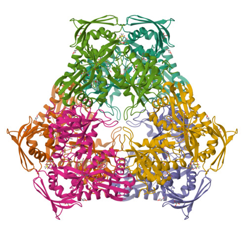
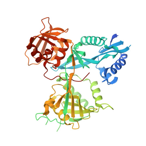
Crystal structure of acetyltransferase Eis from M. tuberculosis in complex with venlafaxine


Experimental Data Snapshot

  • Method: X-RAY DIFFRACTION
  • Resolution: 2.10 Å
  • R-Value Free: 0.193 
  • R-Value Work: 0.167 
  • R-Value Observed: 0.168 

Starting Model: experimental
View more details

wwPDB Validation   3D Report Full Report


Ligand Structure Quality Assessment 

Created with Raphaël 2.3.0Worse 01 BetterLigand structure goodness of fit to experimental dataBest fitted VFXClick on this verticalbar to view details

This is version 1.2 of the entry. See complete history


Literature

Discovery and Mechanistic Analysis of Structurally Diverse Inhibitors of Acetyltransferase Eis among FDA-Approved Drugs.

Pang, A.H.Green, K.D.Punetha, A.Thamban Chandrika, N.Howard, K.C.Garneau-Tsodikova, S.Tsodikov, O.V.

(2023) Biochemistry 62: 710-721

  • DOI: https://doi.org/10.1021/acs.biochem.2c00658
  • Primary Citation of Related Structures:  
    8F4A, 8F4T, 8F4U, 8F4W, 8F4Z, 8F51, 8F55, 8F57, 8F58

  • PubMed Abstract: 

    Over one and a half million people die of tuberculosis (TB) each year. Multidrug-resistant TB infections are especially dangerous, and new drugs are needed to combat them. The high cost and complexity of drug development make repositioning of drugs that are already in clinical use for other indications a potentially time- and money-saving avenue. In this study, we identified among existing drugs five compounds: azelastine, venlafaxine, chloroquine, mefloquine, and proguanil as inhibitors of acetyltransferase Eis from Mycobacterium tuberculosis , a causative agent of TB. Eis upregulation is a cause of clinically relevant resistance of TB to kanamycin, which is inactivated by Eis-catalyzed acetylation. Crystal structures of these drugs as well as chlorhexidine in complexes with Eis showed that these inhibitors were bound in the aminoglycoside binding cavity, consistent with their established modes of inhibition with respect to kanamycin. Among three additionally synthesized compounds, a proguanil analogue, designed based on the crystal structure of the Eis-proguanil complex, was 3-fold more potent than proguanil. The crystal structures of these compounds in complexes with Eis explained their inhibitory potencies. These initial efforts in rational drug repositioning can serve as a starting point in further development of Eis inhibitors.


  • Organizational Affiliation

    Department of Pharmaceutical Sciences, College of Pharmacy, University of Kentucky, 789 South Limestone Street, Lexington, Kentucky40536-0596, United States.


Macromolecules
Find similar proteins by:  (by identity cutoff)  |  3D Structure
Entity ID: 1
MoleculeChains Sequence LengthOrganismDetailsImage
N-acetyltransferase Eis422Mycobacterium tuberculosis H37RvMutation(s): 1 
Gene Names: eisRv2416cMTCY253.04
EC: 2.3.1
UniProt
Find proteins for P9WFK7 (Mycobacterium tuberculosis (strain ATCC 25618 / H37Rv))
Explore P9WFK7 
Go to UniProtKB:  P9WFK7
Entity Groups  
Sequence Clusters30% Identity50% Identity70% Identity90% Identity95% Identity100% Identity
UniProt GroupP9WFK7
Sequence Annotations
Expand
  • Reference Sequence
Small Molecules
Ligands 4 Unique
IDChains Name / Formula / InChI Key2D Diagram3D Interactions
VFX (Subject of Investigation/LOI)
Query on VFX

Download Ideal Coordinates CCD File 
B [auth A]1-[(1R)-2-(dimethylamino)-1-(4-methoxyphenyl)ethyl]cyclohexan-1-ol
C17 H27 N O2
PNVNVHUZROJLTJ-INIZCTEOSA-N
PEG
Query on PEG

Download Ideal Coordinates CCD File 
F [auth A],
I [auth A]
DI(HYDROXYETHYL)ETHER
C4 H10 O3
MTHSVFCYNBDYFN-UHFFFAOYSA-N
GOL
Query on GOL

Download Ideal Coordinates CCD File 
D [auth A]
E [auth A]
G [auth A]
H [auth A]
J [auth A]
GLYCEROL
C3 H8 O3
PEDCQBHIVMGVHV-UHFFFAOYSA-N
DMS
Query on DMS

Download Ideal Coordinates CCD File 
C [auth A]DIMETHYL SULFOXIDE
C2 H6 O S
IAZDPXIOMUYVGZ-UHFFFAOYSA-N
Experimental Data & Validation

Experimental Data

  • Method: X-RAY DIFFRACTION
  • Resolution: 2.10 Å
  • R-Value Free: 0.193 
  • R-Value Work: 0.167 
  • R-Value Observed: 0.168 
  • Space Group: H 3 2
Unit Cell:
Length ( Å )Angle ( ˚ )
a = 175.008α = 90
b = 175.008β = 90
c = 124.366γ = 120
Software Package:
Software NamePurpose
REFMACrefinement
HKL-2000data scaling
HKL-2000data reduction
PHASERphasing

Structure Validation

View Full Validation Report


Ligand Structure Quality Assessment 

Created with Raphaël 2.3.0Worse 01 BetterLigand structure goodness of fit to experimental dataBest fitted VFXClick on this verticalbar to view details

Entry History & Funding Information

Deposition Data


Funding OrganizationLocationGrant Number
National Institutes of Health/National Institute Of Allergy and Infectious Diseases (NIH/NIAID)United StatesAI090048

Revision History  (Full details and data files)

  • Version 1.0: 2023-02-01
    Type: Initial release
  • Version 1.1: 2023-02-22
    Changes: Database references
  • Version 1.2: 2023-10-25
    Changes: Data collection, Refinement description