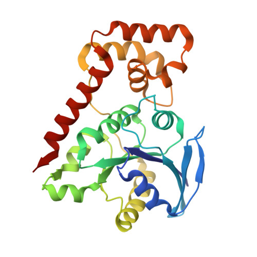
9BA6

High-resolution crystal structure of Vibrio cholerae NFeoB in the apo form

  • Classification: TRANSPORT PROTEIN
  • Organism(s): Vibrio cholerae
  • Expression System: Escherichia coli
  • Mutation(s): No 

  • Deposited: 2024-04-03 Released: 2024-10-30 
  • Deposition Author(s): Lee, M., Smith, A.T.
  • Funding Organization(s): National Institutes of Health/National Institute of General Medical Sciences (NIH/NIGMS), National Science Foundation (NSF, United States)

Experimental Data Snapshot

  • Method: X-RAY DIFFRACTION
  • Resolution: 2.38 Å
  • R-Value Free: 0.267 
  • R-Value Work: 0.205 
  • R-Value Observed: 0.208 

Starting Model: experimental
View more details

wwPDB Validation   3D Report Full Report


This is version 1.0 of the entry. See complete history


Literature

Structural determinants of Vibrio cholerae FeoB nucleotide promiscuity.

Lee, M.Magante, K.Gomez-Garzon, C.Payne, S.M.Smith, A.T.

(2024) J Biol Chem 300: 107663-107663

  • DOI: https://doi.org/10.1016/j.jbc.2024.107663
  • Primary Citation of Related Structures:  
    8VWL, 8VWN, 9BA6, 9BA7

  • PubMed Abstract: 

    Ferrous iron (Fe 2+ ) is required for the growth and virulence of many pathogenic bacteria, including Vibrio cholerae (Vc), the causative agent of the disease cholera. For this bacterium, Feo is the primary system that transports Fe 2+ into the cytosol. FeoB, the main component of this system, is regulated by a soluble cytosolic domain termed NFeoB. Recent reanalysis has shown that NFeoBs can be classified as either GTP-specific or NTP-promiscuous, but the structural and mechanistic bases for these differences were not known. To explore this intriguing property of FeoB, we solved the X-ray crystal structures of VcNFeoB in both the apo and the GDP-bound forms. Surprisingly, this promiscuous NTPase displayed a canonical NFeoB G-protein fold like GTP-specific NFeoBs. Using structural bioinformatics, we hypothesized that residues surrounding the nucleobase could be important for both nucleotide affinity and specificity. We then solved the X-ray crystal structures of N150T VcNFeoB in the apo and GDP-bound forms to reveal H-bonding differences surrounding the guanine nucleobase. Interestingly, isothermal titration calorimetry revealed similar binding thermodynamics of the WT and N150T proteins to guanine nucleotides, while the behavior in the presence of adenine nucleotides was dramatically different. AlphaFold models of VcNFeoB in the presence of ADP and ATP showed important conformational changes that contribute to nucleotide specificity among FeoBs. Combined, these results provide a structural framework for understanding FeoB nucleotide promiscuity, which could be an adaptive measure utilized by pathogens to ensure adequate levels of intracellular iron across multiple metabolic landscapes.


  • Organizational Affiliation

    Department of Chemistry and Biochemistry, University of Maryland, Baltimore County, Baltimore, Maryland, USA.


Macromolecules
Find similar proteins by:  (by identity cutoff)  |  3D Structure
Entity ID: 1
MoleculeChains Sequence LengthOrganismDetailsImage
Ferrous iron transport protein B
A, B
261Vibrio choleraeMutation(s): 0 
Gene Names: feoB_1ERS013165_00117
UniProt
Find proteins for A0A655NVH2 (Vibrio cholerae)
Explore A0A655NVH2 
Go to UniProtKB:  A0A655NVH2
Entity Groups  
Sequence Clusters30% Identity50% Identity70% Identity90% Identity95% Identity100% Identity
UniProt GroupA0A655NVH2
Sequence Annotations
Expand
  • Reference Sequence
Small Molecules
Ligands 2 Unique
IDChains Name / Formula / InChI Key2D Diagram3D Interactions
GOL
Query on GOL

Download Ideal Coordinates CCD File 
C [auth A]
D [auth A]
E [auth A]
F [auth A]
H [auth B]
C [auth A],
D [auth A],
E [auth A],
F [auth A],
H [auth B],
I [auth B],
J [auth B]
GLYCEROL
C3 H8 O3
PEDCQBHIVMGVHV-UHFFFAOYSA-N
CL
Query on CL

Download Ideal Coordinates CCD File 
G [auth A],
K [auth B]
CHLORIDE ION
Cl
VEXZGXHMUGYJMC-UHFFFAOYSA-M
Experimental Data & Validation

Experimental Data

  • Method: X-RAY DIFFRACTION
  • Resolution: 2.38 Å
  • R-Value Free: 0.267 
  • R-Value Work: 0.205 
  • R-Value Observed: 0.208 
  • Space Group: P 1 21 1
Unit Cell:
Length ( Å )Angle ( ˚ )
a = 53.962α = 90
b = 73.442β = 94.25
c = 63.027γ = 90
Software Package:
Software NamePurpose
PHENIXrefinement
xia2data reduction
xia2data scaling
PHASERphasing

Structure Validation

View Full Validation Report


Entry History & Funding Information

Deposition Data


Funding OrganizationLocationGrant Number
National Institutes of Health/National Institute of General Medical Sciences (NIH/NIGMS)United StatesGM133497
National Science Foundation (NSF, United States)United StatesCHE1844624

Revision History  (Full details and data files)

  • Version 1.0: 2024-10-30
    Type: Initial release