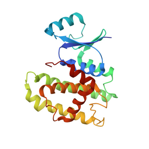
Native and inhibited structure of a Mu class-related glutathione S-transferase from Plasmodium falciparum
Perbandt, M., Burmeister, C., Walter, R.D., Betzel, C., Liebau, E.(2004) J Biol Chem 279: 1336-1342
- PubMed: 12972411
- DOI: https://doi.org/10.1074/jbc.M309663200
- Primary Citation of Related Structures:
1PA3, 1Q4J - PubMed Abstract:
The parasite Plasmodium falciparum causes malaria tropica, the most prevailing parasitic disease worldwide, with 300-500 million infections and 1.5-2.7 million deaths/year. The emergence of strains resistant to drugs used for prophylaxis and treatment and no vaccine available makes the structural analysis of potential drug targets essential. For that reason, we analyzed the three-dimensional structure of the glutathione S-transferase from P. falciparum (Pf-GST1) in the apoform and in complex with its inhibitor S-hexyl-glutathione. The structures have been analyzed to 2.6 and 2.2 A, respectively. Pf-GST1 shares several structural features with the Mu-type GSTs and is therefore closely related to this class, even though alignments with its members display low sequence identities in the range of 20-33%. Upon S-hexyl-glutathione binding, the overall structure and the glutathione-binding site (G-site) remain almost unchanged with the exception of the flexible C terminus. The detailed comparison of the parasitic enzyme with the human host Mu-class enzyme reveals that, although the overall structure is homologue, the shape of the hydrophobic binding pocket (H-site) differs substantially. In the human enzyme, it is shielded from one side by the large Mu-loop, whereas in Pf-GST1 the Mu-loop is truncated and the space to recognize and bind voluminous substrates is extended. This structural feature can be exploited to support the design of specific and parasite-selective inhibitors.
Organizational Affiliation:
Department of Biochemistry and Molecular Biology, University of Hamburg, Martin Luther King Platz 6, 20146 Hamburg, Germany. [email protected]


