RCSB PDB - 2GJ5: Crystal structure of a secondary vitamin D3 binding site of milk beta-lactoglobulin

 2GJ5

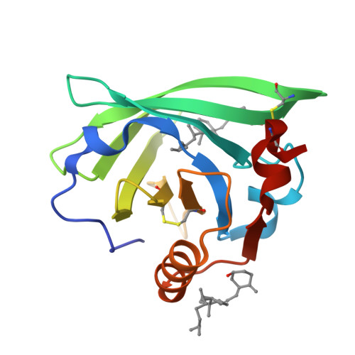
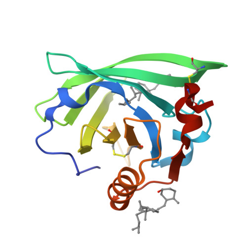
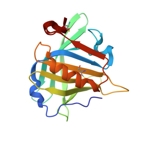
Crystal structure of a secondary vitamin D3 binding site of milk beta-lactoglobulin


Experimental Data Snapshot

  • Method: X-RAY DIFFRACTION
  • Resolution: 2.40 Å
  • R-Value Free: 0.298 
  • R-Value Work: 0.236 
  • R-Value Observed: 0.241 

Starting Model: experimental
View more details

wwPDB Validation   3D Report Full Report


Ligand Structure Quality Assessment 

Created with Raphaël 2.3.0Worse 01 BetterLigand structure goodness of fit to experimental dataBest fitted VD3Click on this verticalbar to view details

This is version 1.3 of the entry. See complete history


Literature

Crystal structure of a secondary vitamin D3 binding site of milk beta-lactoglobulin.

Yang, M.C.Guan, H.H.Liu, M.Y.Lin, Y.H.Yang, J.M.Chen, W.L.Chen, C.J.Mao, S.J.

(2008) Proteins 71: 1197-1210

  • DOI: https://doi.org/10.1002/prot.21811
  • Primary Citation of Related Structures:  
    2GJ5

  • PubMed Abstract: 

    Beta-lactoglobulin (beta-LG), one of the most investigated proteins, is a major bovine milk protein with a predominantly beta structure. The structural function of the only alpha-helix with three turns at the C-terminus is unknown. Vitamin D(3) binds to the central calyx formed by the beta-strands. Whether there are two vitamin D binding-sites in each beta-LG molecule has been a subject of controversy. Here, we report a second vitamin D(3) binding site identified by synchrotron X-ray diffraction (at 2.4 A resolution). In the central calyx binding mode, the aliphatic tail of vitamin D(3) clearly inserts into the binding cavity, where the 3-OH group of vitamin D(3) binds externally. The electron density map suggests that the 3-OH group interacts with the carbonyl of Lys-60 forming a hydrogen bond (2.97 A). The second binding site, however, is near the surface at the C-terminus (residues 136-149) containing part of an alpha-helix and a beta-strand I with 17.91 A in length, while the span of vitamin D(3) is about 12.51 A. A remarkable feature of the second exosite is that it combines an amphipathic alpha-helix providing nonpolar residues (Phe-136, Ala-139, and Leu-140) and a beta-strand providing a nonpolar (Ile-147) and a buried polar residue (Arg-148). They are linked by a hydrophobic loop (Ala-142, Leu-143, Pro-144, and Met-145). Thus, the binding pocket furnishes strong hydrophobic force to stabilize vitamin D(3) binding. This finding provides a new insight into the interaction between vitamin D(3) and beta-LG, in which the exosite may provide another route for the transport of vitamin D(3) in vitamin D(3) fortified dairy products. Atomic coordinates for the crystal structure of beta-LG-vitamin D(3) complex described in this work have been deposited in the PDB (access code 2GJ5).


  • Organizational Affiliation

    Department of Biological Science and Technology, College of Biological Science and Technology, National Chiao Tung University, Taiwan, Republic of China.


Macromolecules
Find similar proteins by:  (by identity cutoff)  |  3D Structure
Entity ID: 1
MoleculeChains Sequence LengthOrganismDetailsImage
Beta-lactoglobulin162Bos taurusMutation(s): 0 
UniProt
Find proteins for P02754 (Bos taurus)
Explore P02754 
Go to UniProtKB:  P02754
Entity Groups  
Sequence Clusters30% Identity50% Identity70% Identity90% Identity95% Identity100% Identity
UniProt GroupP02754
Sequence Annotations
Expand
  • Reference Sequence
Small Molecules
Ligands 1 Unique
IDChains Name / Formula / InChI Key2D Diagram3D Interactions
VD3
Query on VD3

Download Ideal Coordinates CCD File 
B [auth A],
C [auth A]
(1S,3Z)-3-[(2E)-2-[(1R,3AR,7AS)-7A-METHYL-1-[(2R)-6-METHYLHEPTAN-2-YL]-2,3,3A,5,6,7-HEXAHYDRO-1H-INDEN-4-YLIDENE]ETHYLI DENE]-4-METHYLIDENE-CYCLOHEXAN-1-OL
C27 H44 O
QYSXJUFSXHHAJI-RWDMXNMGSA-N
Binding Affinity Annotations 
IDSourceBinding Affinity
VD3 PDBBind:  2GJ5 Kd: 4.74 (nM) from 1 assay(s)
Experimental Data & Validation

Experimental Data

  • Method: X-RAY DIFFRACTION
  • Resolution: 2.40 Å
  • R-Value Free: 0.298 
  • R-Value Work: 0.236 
  • R-Value Observed: 0.241 
  • Space Group: P 32 2 1
Unit Cell:
Length ( Å )Angle ( ˚ )
a = 53.782α = 90
b = 53.782β = 90
c = 111.578γ = 120
Software Package:
Software NamePurpose
ADSCdata collection
HKL-2000data reduction
CNSrefinement
HKL-2000data scaling
CNSphasing

Structure Validation

View Full Validation Report


Ligand Structure Quality Assessment 

Created with Raphaël 2.3.0Worse 01 BetterLigand structure goodness of fit to experimental dataBest fitted VD3Click on this verticalbar to view details

Entry History 

Deposition Data

Revision History  (Full details and data files)

  • Version 1.0: 2007-10-02
    Type: Initial release
  • Version 1.1: 2011-07-13
    Changes: Version format compliance
  • Version 1.2: 2023-10-25
    Changes: Data collection, Database references, Derived calculations, Refinement description, Structure summary
  • Version 1.3: 2024-10-23
    Changes: Structure summary