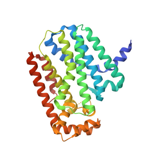
2Q80

Crystal structure of human geranylgeranyl pyrophosphate synthase bound to GGPP


Experimental Data Snapshot

  • Method: X-RAY DIFFRACTION
  • Resolution: 2.70 Å
  • R-Value Free: 0.253 
  • R-Value Work: 0.202 
  • R-Value Observed: 0.204 

Starting Model: experimental
View more details

wwPDB Validation   3D Report Full Report


Ligand Structure Quality Assessment 


This is version 1.5 of the entry. See complete history


Literature

The crystal structure of human geranylgeranyl pyrophosphate synthase reveals a novel hexameric arrangement and inhibitory product binding

Kavanagh, K.L.Dunford, J.E.Bunkoczi, G.Russell, R.G.Oppermann, U.

(2006) J Biol Chem 281: 22004-22012

  • DOI: https://doi.org/10.1074/jbc.M602603200
  • Primary Citation of Related Structures:  
    2Q80

  • PubMed Abstract: 

    Modification of GTPases with isoprenoid molecules derived from geranylgeranyl pyrophosphate or farnesyl pyrophosphate is an essential requisite for cellular signaling pathways. The synthesis of these isoprenoids proceeds in mammals through the mevalonate pathway, and the final steps in the synthesis are catalyzed by the related enzymes farnesyl pyrophosphate synthase and geranylgeranyl pyrophosphate synthase. Both enzymes play crucial roles in cell survival, and inhibition of farnesyl pyrophosphate synthase by nitrogen-containing bisphosphonates is an established concept in the treatment of bone disorders such as osteoporosis or certain forms of cancer in bone. Here we report the crystal structure of human geranylgeranyl pyrophosphate synthase, the first mammalian ortholog to have its x-ray structure determined. It reveals that three dimers join together to form a propeller-bladed hexameric molecule with a mass of approximately 200 kDa. Structure-based sequence alignments predict this quaternary structure to be restricted to mammalian and insect orthologs, whereas fungal, bacterial, archaeal, and plant forms exhibit the dimeric organization also observed in farnesyl pyrophosphate synthase. Geranylgeranyl pyrophosphate derived from heterologous bacterial expression is tightly bound in a cavity distinct from the chain elongation site described for farnesyl pyrophosphate synthase. The structure most likely represents an inhibitory complex, which is further corroborated by steady-state kinetics, suggesting a possible feedback mechanism for regulating enzyme activity. Structural comparisons between members of this enzyme class give deeper insights into conserved features important for catalysis.


  • Organizational Affiliation

    Structural Genomics Consortium, Botnar Research Centre, University of Oxford, Oxford OX3 7LD, United Kingdom. Electronic address: [email protected].


Macromolecules
Find similar proteins by:  (by identity cutoff)  |  3D Structure
Entity ID: 1
MoleculeChains Sequence LengthOrganismDetailsImage
Geranylgeranyl pyrophosphate synthetase
A, B, C, D, E
A, B, C, D, E, F
301Homo sapiensMutation(s): 0 
Gene Names: GGPS1
EC: 2.5.1.1 (PDB Primary Data), 2.5.1.10 (PDB Primary Data), 2.5.1.29 (PDB Primary Data), 2.5.1 (UniProt)
UniProt & NIH Common Fund Data Resources
Find proteins for O95749 (Homo sapiens)
Explore O95749 
Go to UniProtKB:  O95749
PHAROS:  O95749
GTEx:  ENSG00000152904 
Entity Groups  
Sequence Clusters30% Identity50% Identity70% Identity90% Identity95% Identity100% Identity
UniProt GroupO95749
Sequence Annotations
Expand
  • Reference Sequence
Small Molecules
Ligands 2 Unique
IDChains Name / Formula / InChI Key2D Diagram3D Interactions
GRG
Query on GRG

Download Ideal Coordinates CCD File 
I [auth A]
L [auth B]
O [auth C]
R [auth D]
U [auth E]
I [auth A],
L [auth B],
O [auth C],
R [auth D],
U [auth E],
X [auth F]
GERANYLGERANYL DIPHOSPHATE
C20 H36 O7 P2
OINNEUNVOZHBOX-QIRCYJPOSA-N
MG
Query on MG

Download Ideal Coordinates CCD File 
G [auth A]
H [auth A]
J [auth B]
K [auth B]
M [auth C]
G [auth A],
H [auth A],
J [auth B],
K [auth B],
M [auth C],
N [auth C],
P [auth D],
Q [auth D],
S [auth E],
T [auth E],
V [auth F],
W [auth F]
MAGNESIUM ION
Mg
JLVVSXFLKOJNIY-UHFFFAOYSA-N
Binding Affinity Annotations 
IDSourceBinding Affinity
GRG PDBBind:  2Q80 Ki: 2.50e+4 (nM) from 1 assay(s)
Experimental Data & Validation

Experimental Data

  • Method: X-RAY DIFFRACTION
  • Resolution: 2.70 Å
  • R-Value Free: 0.253 
  • R-Value Work: 0.202 
  • R-Value Observed: 0.204 
  • Space Group: P 41 21 2
Unit Cell:
Length ( Å )Angle ( ˚ )
a = 141.248α = 90
b = 141.248β = 90
c = 211.705γ = 90
Software Package:
Software NamePurpose
PHASERphasing
REFMACrefinement
XDSdata reduction
XSCALEdata scaling

Structure Validation

View Full Validation Report


Ligand Structure Quality Assessment 


Entry History 

Deposition Data

Revision History  (Full details and data files)

  • Version 1.0: 2007-06-19
    Type: Initial release
  • Version 1.1: 2008-05-01
    Changes: Version format compliance
  • Version 1.2: 2011-07-13
    Changes: Advisory, Version format compliance
  • Version 1.3: 2012-12-05
    Changes: Structure summary
  • Version 1.4: 2024-04-03
    Changes: Data collection, Database references, Derived calculations, Refinement description
  • Version 1.5: 2024-12-25
    Changes: Advisory, Derived calculations, Structure summary