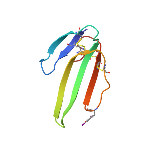
Different Interactions between Mt7 Toxin and the Human Muscarinic M1 Receptor in its Free and N-Methylscopolamine-Occupied States.
Fruchart-Gaillard, C., Mourier, G., Marquer, C., Stura, E., Birdsall, N.J.M., Servent, D.(2008) Mol Pharmacol 74: 1554
- PubMed: 18784346
- DOI: https://doi.org/10.1124/mol.108.050773
- Primary Citation of Related Structures:
2VLW - PubMed Abstract:
Muscarinic MT7 toxin is a highly selective and potent antagonist of the M(1) subtype of muscarinic receptor and acts by binding to an allosteric site. To identify the molecular determinants by which MT7 toxin interacts with this receptor in its free and NMS-occupied states, the effect on toxin potency of alanine substitution was evaluated in equilibrium and kinetic binding experiments as well as in functional assays. The determination of the crystallographic structure of an MT7-derivative (MT7-diiodoTyr51) allowed the selection of candidate residues that are accessible and present on both faces of the three toxin loops. The equilibrium binding data are consistent with negative cooperativity between N-methylscopolamine (NMS) and wild-type or modified MT7 and highlight the critical role of the tip of the central loop of the toxin (Arg34, Met35 Tyr36) in its interaction with the unoccupied receptor. Examination of the potency of wild-type and modified toxins to allosterically decrease the dissociation rate of [(3)H]NMS allowed the identification of the MT7 residues involved in its interaction with the NMS-occupied receptor. In contrast to the results with the unoccupied receptor, the most important residue for this interaction was Tyr36 in loop II, assisted by Trp10 in loop I and Arg52 in loop III. The critical role of the tips of the MT7 loops was also confirmed in functional experiments. The high specificity of the MT7-M(1) receptor interaction exploits several MT7-specific residues and reveals a different mode of interaction of the toxin with the free and NMS-occupied states of the receptor.
Organizational Affiliation:
Commissariat à l'Energie Atomique, Institut de Biologie et de Tecnologies de Saclay, Service d'Ingénierie Moléculaire des Protéines, Laboratoire de Toxinologie Moléculaire et Biotechnologie, Gif sur Yvette, France.


