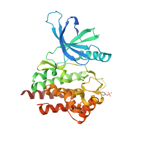
3LXN

Structural and Thermodynamic Characterization of the TYK2 and JAK3 Kinase Domains in Complex with CP-690550 and CMP-6


Experimental Data Snapshot

  • Method: X-RAY DIFFRACTION
  • Resolution: 2.50 Å
  • R-Value Free: 0.286 
  • R-Value Work: 0.220 
  • R-Value Observed: 0.223 

Starting Model: experimental
View more details

wwPDB Validation   3D Report Full Report


Ligand Structure Quality Assessment 


This is version 1.5 of the entry. See complete history


Literature

Structural and thermodynamic characterization of the TYK2 and JAK3 kinase domains in complex with CP-690550 and CMP-6.

Chrencik, J.E.Patny, A.Leung, I.K.Korniski, B.Emmons, T.L.Hall, T.Weinberg, R.A.Gormley, J.A.Williams, J.M.Day, J.E.Hirsch, J.L.Kiefer, J.R.Leone, J.W.Fischer, H.D.Sommers, C.D.Huang, H.C.Jacobsen, E.J.Tenbrink, R.E.Tomasselli, A.G.Benson, T.E.

(2010) J Mol Biol 400: 413-433

  • DOI: https://doi.org/10.1016/j.jmb.2010.05.020
  • Primary Citation of Related Structures:  
    3LXK, 3LXL, 3LXN, 3LXP

  • PubMed Abstract: 

    Janus kinases (JAKs) are critical regulators of cytokine pathways and attractive targets of therapeutic value in both inflammatory and myeloproliferative diseases. Although the crystal structures of active JAK1 and JAK2 kinase domains have been reported recently with the clinical compound CP-690550, the structures of both TYK2 and JAK3 with CP-690550 have remained outstanding. Here, we report the crystal structures of TYK2, a first in class structure, and JAK3 in complex with PAN-JAK inhibitors CP-690550 ((3R,4R)-3-[4-methyl-3-[N-methyl-N-(7H-pyrrolo[2,3-d]pyrimidin-4-yl)amino]piperidin-1-yl]-3-oxopropionitrile) and CMP-6 (tetracyclic pyridone 2-t-butyl-9-fluoro-3,6-dihydro-7H-benz[h]-imidaz[4,5-f]isoquinoline-7-one), both of which bind in the ATP-binding cavities of both JAK isozymes in orientations similar to that observed in crystal structures of JAK1 and JAK2. Additionally, a complete thermodynamic characterization of JAK/CP-690550 complex formation was completed by isothermal titration calorimetry, indicating the critical role of the nitrile group from the CP-690550 compound. Finally, computational analysis using WaterMap further highlights the critical positioning of the CP-690550 nitrile group in the displacement of an unfavorable water molecule beneath the glycine-rich loop. Taken together, the data emphasize the outstanding properties of the kinome-selective JAK inhibitor CP-690550, as well as the challenges in obtaining JAK isozyme-selective inhibitors due to the overall structural and sequence similarities between the TYK2, JAK1, JAK2 and JAK3 isozymes. Nevertheless, subtle amino acid variations of residues lining the ligand-binding cavity of the JAK enzymes, as well as the global positioning of the glycine-rich loop, might provide the initial clues to obtaining JAK-isozyme selective inhibitors.


  • Organizational Affiliation

    Pfizer Inc. 700 Chesterfield Parkway West, Chesterfield, MO 63017, USA. [email protected]


Macromolecules
Find similar proteins by:  (by identity cutoff)  |  3D Structure
Entity ID: 1
MoleculeChains Sequence LengthOrganismDetailsImage
Non-receptor tyrosine-protein kinase TYK2318Homo sapiensMutation(s): 5 
Gene Names: TYK2
EC: 2.7.10.2
UniProt & NIH Common Fund Data Resources
Find proteins for P29597 (Homo sapiens)
Explore P29597 
Go to UniProtKB:  P29597
PHAROS:  P29597
GTEx:  ENSG00000105397 
Entity Groups  
Sequence Clusters30% Identity50% Identity70% Identity90% Identity95% Identity100% Identity
UniProt GroupP29597
Sequence Annotations
Expand
  • Reference Sequence
Small Molecules
Ligands 1 Unique
IDChains Name / Formula / InChI Key2D Diagram3D Interactions
MI1
Query on MI1

Download Ideal Coordinates CCD File 
B [auth A]3-{(3R,4R)-4-methyl-3-[methyl(7H-pyrrolo[2,3-d]pyrimidin-4-yl)amino]piperidin-1-yl}-3-oxopropanenitrile
C16 H20 N6 O
UJLAWZDWDVHWOW-YPMHNXCESA-N
Modified Residues  1 Unique
IDChains TypeFormula2D DiagramParent
PTR
Query on PTR
A
L-PEPTIDE LINKINGC9 H12 N O6 PTYR
Binding Affinity Annotations 
IDSourceBinding Affinity
MI1 BindingDB:  3LXN Ki: min: 4.39, max: 7.8 (nM) from 3 assay(s)
Kd: min: 4.8, max: 1.00e+4 (nM) from 4 assay(s)
IC50: min: 0.4, max: 1200 (nM) from 35 assay(s)
EC50: 5 (nM) from 1 assay(s)
Experimental Data & Validation

Experimental Data

  • Method: X-RAY DIFFRACTION
  • Resolution: 2.50 Å
  • R-Value Free: 0.286 
  • R-Value Work: 0.220 
  • R-Value Observed: 0.223 
  • Space Group: P 21 21 21
Unit Cell:
Length ( Å )Angle ( ˚ )
a = 36.306α = 90
b = 74.262β = 90
c = 106.46γ = 90
Software Package:
Software NamePurpose
HKL-2000data collection
AMoREphasing
REFMACrefinement
HKL-2000data reduction
HKL-2000data scaling

Structure Validation

View Full Validation Report


Ligand Structure Quality Assessment 


Entry History 

Deposition Data

Revision History  (Full details and data files)

  • Version 1.0: 2010-06-02
    Type: Initial release
  • Version 1.1: 2011-07-13
    Changes: Version format compliance
  • Version 1.2: 2021-10-13
    Changes: Database references, Derived calculations
  • Version 1.3: 2023-09-06
    Changes: Data collection, Refinement description
  • Version 1.4: 2023-11-22
    Changes: Data collection
  • Version 1.5: 2024-10-09
    Changes: Structure summary