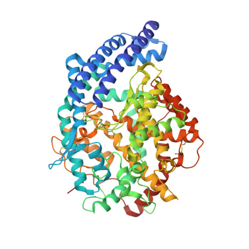
4BZR

Human testis angiotensin converting enzyme in complex with K-26


Experimental Data Snapshot

  • Method: X-RAY DIFFRACTION
  • Resolution: 1.84 Å
  • R-Value Free: 0.252 
  • R-Value Work: 0.214 
  • R-Value Observed: 0.216 

Starting Model: experimental
View more details

wwPDB Validation   3D Report Full Report


Ligand Structure Quality Assessment 


This is version 2.2 of the entry. See complete history


Literature

Interkingdom Pharmacology of Angiotensin-I Converting Enzyme Inhibitor Phosphonates Produced by Actinomycetes

Kramer, G.J.Mohd, A.Schwager, S.L.U.Masuyer, G.Acharya, K.R.Sturrock, E.D.Bachmann, B.O.

(2014) ACS Med Chem Lett 5: 346

  • DOI: https://doi.org/10.1021/ml4004588
  • Primary Citation of Related Structures:  
    4BZR, 4BZS

  • PubMed Abstract: 

    The K-26 family of bacterial secondary metabolites are N-modified tripeptides terminated by an unusual phosphonate analog of tyrosine. These natural products, produced via three different actinomycetales, are potent inhibitors of human angiotensin-I converting enzyme (ACE). Herein we investigate the interkingdom pharmacology of the K-26 family by synthesizing these metabolites and assessing their potency as inhibitors of both the N-terminal and C-terminal domains of human ACE. In most cases, selectivity for the C-terminal domain of ACE is displayed. Co-crystallization of K-26 in both domains of human ACE reveals the structural basis of the potent inhibition and has shown an unusual binding motif that may guide future design of domain-selective inhibitors. Finally, the activity of K-26 is assayed against a cohort of microbially produced ACE relatives. In contrast to the synthetic ACE inhibitor captopril, which demonstrates broad interkingdom inhibition of ACE-like enzymes, K-26 selectively targets the eukaryotic family.


  • Organizational Affiliation

    Vanderbilt University Department of Chemistry, 7300 Stevenson Center, Nashville, Tennessee 37204, United States.


Macromolecules
Find similar proteins by:  (by identity cutoff)  |  3D Structure
Entity ID: 1
MoleculeChains Sequence LengthOrganismDetailsImage
ANGIOTENSIN-CONVERTING ENZYME589Homo sapiensMutation(s): 0 
EC: 3.2.1 (PDB Primary Data), 3.4.15.1 (PDB Primary Data)
UniProt & NIH Common Fund Data Resources
Find proteins for P12821 (Homo sapiens)
Explore P12821 
Go to UniProtKB:  P12821
PHAROS:  P12821
GTEx:  ENSG00000159640 
Entity Groups  
Sequence Clusters30% Identity50% Identity70% Identity90% Identity95% Identity100% Identity
UniProt GroupP12821
Glycosylation
Glycosylation Sites: 2Go to GlyGen: P12821-1
Sequence Annotations
Expand
  • Reference Sequence
Oligosaccharides

Help

Entity ID: 2
MoleculeChains Length2D Diagram Glycosylation3D Interactions
beta-D-mannopyranose-(1-4)-2-acetamido-2-deoxy-beta-D-glucopyranose-(1-4)-[alpha-L-fucopyranose-(1-6)]2-acetamido-2-deoxy-beta-D-glucopyranose
B
4N-Glycosylation
Glycosylation Resources
GlyTouCan:  G32152BH
GlyCosmos:  G32152BH
GlyGen:  G32152BH
Small Molecules
Ligands 6 Unique
IDChains Name / Formula / InChI Key2D Diagram3D Interactions
K26
Query on K26

Download Ideal Coordinates CCD File 
E [auth A]N-ACETYL-L-ILE-L-TYR-(R)-1-AMINO-2-(4-HYDROXYPHENYL)ETHYLPHOSPHONIC ACID
C25 H34 N3 O8 P
ZFRNBYWFOLDQKG-FDMHNHSTSA-N
PE4
Query on PE4

Download Ideal Coordinates CCD File 
D [auth A]2-{2-[2-(2-{2-[2-(2-ETHOXY-ETHOXY)-ETHOXY]-ETHOXY}-ETHOXY)-ETHOXY]-ETHOXY}-ETHANOL
C16 H34 O8
PJWQOENWHPEPKI-UHFFFAOYSA-N
NAG
Query on NAG

Download Ideal Coordinates CCD File 
I [auth A]2-acetamido-2-deoxy-beta-D-glucopyranose
C8 H15 N O6
OVRNDRQMDRJTHS-FMDGEEDCSA-N
ZN
Query on ZN

Download Ideal Coordinates CCD File 
H [auth A]ZINC ION
Zn
PTFCDOFLOPIGGS-UHFFFAOYSA-N
ACT
Query on ACT

Download Ideal Coordinates CCD File 
C [auth A]ACETATE ION
C2 H3 O2
QTBSBXVTEAMEQO-UHFFFAOYSA-M
CL
Query on CL

Download Ideal Coordinates CCD File 
F [auth A],
G [auth A]
CHLORIDE ION
Cl
VEXZGXHMUGYJMC-UHFFFAOYSA-M
Binding Affinity Annotations 
IDSourceBinding Affinity
K26 BindingDB:  4BZR Ki: min: 11, max: 75 (nM) from 2 assay(s)
IC50: min: 11, max: 110 (nM) from 3 assay(s)
Experimental Data & Validation

Experimental Data

  • Method: X-RAY DIFFRACTION
  • Resolution: 1.84 Å
  • R-Value Free: 0.252 
  • R-Value Work: 0.214 
  • R-Value Observed: 0.216 
  • Space Group: P 21 21 21
Unit Cell:
Length ( Å )Angle ( ˚ )
a = 56.25α = 90
b = 84.505β = 90
c = 133.481γ = 90
Software Package:
Software NamePurpose
REFMACrefinement
HKL-2000data reduction
SCALEPACKdata scaling
PHASERphasing

Structure Validation

View Full Validation Report


Ligand Structure Quality Assessment 


Entry History 

Deposition Data

Revision History  (Full details and data files)

  • Version 1.0: 2014-02-26
    Type: Initial release
  • Version 1.1: 2014-06-18
    Changes: Database references
  • Version 2.0: 2020-07-29
    Type: Remediation
    Reason: Carbohydrate remediation
    Changes: Advisory, Atomic model, Data collection, Derived calculations, Other, Structure summary
  • Version 2.1: 2023-12-20
    Changes: Data collection, Database references, Refinement description, Structure summary
  • Version 2.2: 2024-10-23
    Changes: Structure summary