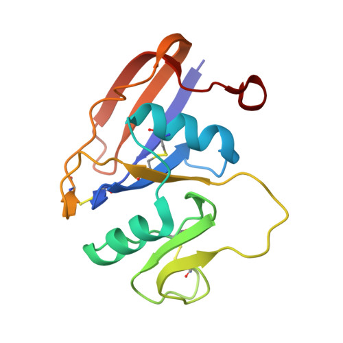
4MRG

Crystal structure of the murine cd44 hyaluronan binding domain complex with a small molecule


Experimental Data Snapshot

  • Method: X-RAY DIFFRACTION
  • Resolution: 1.69 Å
  • R-Value Free: 0.206 
  • R-Value Work: 0.160 
  • R-Value Observed: 0.163 

Starting Model: experimental
View more details

wwPDB Validation   3D Report Full Report


This is version 1.2 of the entry. See complete history


Literature

Fragment-Based Identification of an Inducible Binding Site on Cell Surface Receptor CD44 for the Design of Protein-Carbohydrate Interaction Inhibitors.

Liu, L.K.Finzel, B.C.

(2014) J Med Chem 57: 2714-2725

  • DOI: https://doi.org/10.1021/jm5000276
  • Primary Citation of Related Structures:  
    4MRD, 4MRE, 4MRF, 4MRG, 4MRH, 4NP2, 4NP3

  • PubMed Abstract: 

    Selective inhibitors of hyaluronan (HA) binding to the cell surface receptor CD44 will have value as probes of CD44-mediated signaling and have potential as therapeutic agents in chronic inflammation, cardiovascular disease, and cancer. Using biophysical binding assays, fragment screening, and crystallographic characterization of complexes with the CD44 HA binding domain, we have discovered an inducible pocket adjacent to the HA binding groove into which small molecules may bind. Iterations of fragment combination and structure-driven design have allowed identification of a series of 1,2,3,4-tetrahydroisoquinolines as the first nonglycosidic inhibitors of the CD44-HA interaction. The affinity of these molecules for the CD44 HA binding domain parallels their ability to interfere with CD44 binding to polymeric HA in vitro. X-ray crystallographic complexes of lead compounds are described and compared to a new complex with a short HA tetrasaccharide, to establish the tetrahydroisoquinoline pharmacophore as an attractive starting point for lead optimization.


  • Organizational Affiliation

    Department of Medicinal Chemistry, University of Minnesota , 308 Harvard Street SE, Minneapolis, Minnesota 55455, United States.


Macromolecules
Find similar proteins by:  (by identity cutoff)  |  3D Structure
Entity ID: 1
MoleculeChains Sequence LengthOrganismDetailsImage
CD44 antigen150Mus musculusMutation(s): 0 
Gene Names: Cd44Cd44 Ly-24Ly-24
UniProt
Find proteins for P15379 (Mus musculus)
Explore P15379 
Go to UniProtKB:  P15379
Entity Groups  
Sequence Clusters30% Identity50% Identity70% Identity90% Identity95% Identity100% Identity
UniProt GroupP15379
Sequence Annotations
Expand
  • Reference Sequence
Small Molecules
Ligands 4 Unique
IDChains Name / Formula / InChI Key2D Diagram3D Interactions
24W
Query on 24W

Download Ideal Coordinates CCD File 
B [auth A]1,2,3,4-tetrahydroisoquinolin-5-amine
C9 H12 N2
ZSHCQIHYOFRGNI-UHFFFAOYSA-N
SO4
Query on SO4

Download Ideal Coordinates CCD File 
F [auth A]SULFATE ION
O4 S
QAOWNCQODCNURD-UHFFFAOYSA-L
GOL
Query on GOL

Download Ideal Coordinates CCD File 
D [auth A],
E [auth A]
GLYCEROL
C3 H8 O3
PEDCQBHIVMGVHV-UHFFFAOYSA-N
DMS
Query on DMS

Download Ideal Coordinates CCD File 
C [auth A]DIMETHYL SULFOXIDE
C2 H6 O S
IAZDPXIOMUYVGZ-UHFFFAOYSA-N
Binding Affinity Annotations 
IDSourceBinding Affinity
24W BindingDB:  4MRG Kd: min: 9.00e+5, max: 1.10e+6 (nM) from 2 assay(s)
IC50: 6.70e+6 (nM) from 1 assay(s)
Experimental Data & Validation

Experimental Data

  • Method: X-RAY DIFFRACTION
  • Resolution: 1.69 Å
  • R-Value Free: 0.206 
  • R-Value Work: 0.160 
  • R-Value Observed: 0.163 
  • Space Group: P 1 21 1
Unit Cell:
Length ( Å )Angle ( ˚ )
a = 30.85α = 90
b = 81.58β = 117.93
c = 32.13γ = 90
Software Package:
Software NamePurpose
XSCALEdata scaling
PHASERphasing
REFMACrefinement
PDB_EXTRACTdata extraction
Blu-Icedata collection

Structure Validation

View Full Validation Report


Entry History 

Deposition Data

Revision History  (Full details and data files)

  • Version 1.0: 2014-04-16
    Type: Initial release
  • Version 1.1: 2023-09-20
    Changes: Data collection, Database references, Derived calculations, Refinement description
  • Version 1.2: 2024-11-20
    Changes: Structure summary