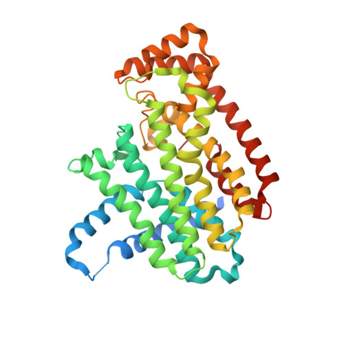
Crystallographic and thermodynamic characterization of phenylaminopyridine bisphosphonates binding to human farnesyl pyrophosphate synthase.
Park, J., Rodionov, D., De Schutter, J.W., Lin, Y.S., Tsantrizos, Y.S., Berghuis, A.M.(2017) PLoS One 12: e0186447-e0186447
- PubMed: 29036218
- DOI: https://doi.org/10.1371/journal.pone.0186447
- Primary Citation of Related Structures:
4NFI, 4NFJ, 4NFK, 4PVX, 4PVY - PubMed Abstract:
Human farnesyl pyrophosphate synthase (hFPPS) catalyzes the production of the 15-carbon isoprenoid farnesyl pyrophosphate. The enzyme is a key regulator of the mevalonate pathway and a well-established drug target. Notably, it was elucidated as the molecular target of nitrogen-containing bisphosphonates, a class of drugs that have been widely successful against bone resorption disorders. More recently, research has focused on the anticancer effects of these inhibitors. In order to achieve increased non-skeletal tissue exposure, we created phenylaminopyridine bisphosphonates (PNP-BPs) that have bulky hydrophobic side chains through a structure-based approach. Some of these compounds have proven to be more potent than the current clinical drugs in a number of antiproliferation assays using multiple myeloma cell lines. In the present work, we characterized the binding of our most potent PNP-BPs to the target enzyme, hFPPS. Co-crystal structures demonstrate that the molecular interactions designed to elicit tighter binding are indeed established. We carried out thermodynamic studies as well; the newly introduced protein-ligand interactions are clearly reflected in the enthalpy of binding measured, which is more favorable for the new PNP-BPs than for the lead compound. These studies also indicate that the affinity of the PNP-BPs to hFPPS is comparable to that of the current drug risedronate. Risedronate forms additional polar interactions via its hydroxyl functional group and thus exhibits more favorable binding enthalpy; however, the entropy of binding is more favorable for the PNP-BPs, owing to the greater desolvation effects resulting from their large hydrophobic side chains. These results therefore confirm the overall validity of our drug design strategy. With a distinctly different molecular scaffold, the PNP-BPs described in this report represent an interesting new group of future drug candidates. Further investigation should follow to characterize the tissue distribution profile and assess the potential clinical benefits of these compounds.
Organizational Affiliation:
Department of Biochemistry, McGill University, Montreal, Quebec, Canada.


