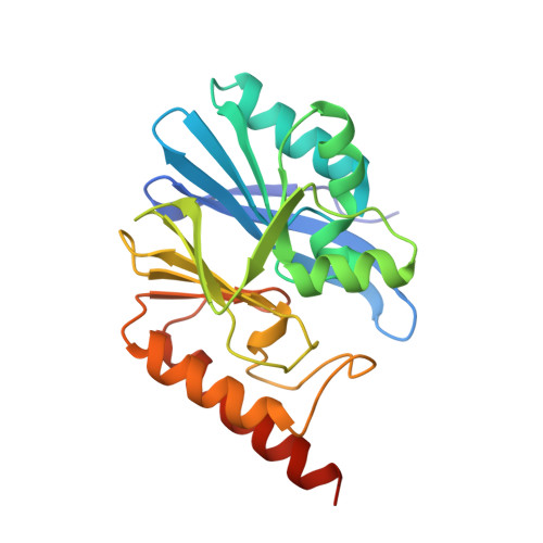
5EV6

Crystal structure of the native, di-zinc metallo-beta-lactamase IMP-1


Experimental Data Snapshot

  • Method: X-RAY DIFFRACTION
  • Resolution: 1.98 Å
  • R-Value Free: 0.204 
  • R-Value Work: 0.172 
  • R-Value Observed: 0.174 

Starting Model: experimental
View more details

wwPDB Validation   3D Report Full Report


This is version 1.3 of the entry. See complete history


Literature

Cross-class metallo-beta-lactamase inhibition by bisthiazolidines reveals multiple binding modes.

Hinchliffe, P.Gonzalez, M.M.Mojica, M.F.Gonzalez, J.M.Castillo, V.Saiz, C.Kosmopoulou, M.Tooke, C.L.Llarrull, L.I.Mahler, G.Bonomo, R.A.Vila, A.J.Spencer, J.

(2016) Proc Natl Acad Sci U S A 113: E3745-E3754

  • DOI: https://doi.org/10.1073/pnas.1601368113
  • Primary Citation of Related Structures:  
    4NQ6, 5EV6, 5EV8, 5EVB, 5EVD, 5EVK, 5EW0, 5EWA

  • PubMed Abstract: 

    Metallo-β-lactamases (MBLs) hydrolyze almost all β-lactam antibiotics and are unaffected by clinically available β-lactamase inhibitors (βLIs). Active-site architecture divides MBLs into three classes (B1, B2, and B3), complicating development of βLIs effective against all enzymes. Bisthiazolidines (BTZs) are carboxylate-containing, bicyclic compounds, considered as penicillin analogs with an additional free thiol. Here, we show both l- and d-BTZ enantiomers are micromolar competitive βLIs of all MBL classes in vitro, with Kis of 6-15 µM or 36-84 µM for subclass B1 MBLs (IMP-1 and BcII, respectively), and 10-12 µM for the B3 enzyme L1. Against the B2 MBL Sfh-I, the l-BTZ enantiomers exhibit 100-fold lower Kis (0.26-0.36 µM) than d-BTZs (26-29 µM). Importantly, cell-based time-kill assays show BTZs restore β-lactam susceptibility of Escherichia coli-producing MBLs (IMP-1, Sfh-1, BcII, and GOB-18) and, significantly, an extensively drug-resistant Stenotrophomonas maltophilia clinical isolate expressing L1. BTZs therefore inhibit the full range of MBLs and potentiate β-lactam activity against producer pathogens. X-ray crystal structures reveal insights into diverse BTZ binding modes, varying with orientation of the carboxylate and thiol moieties. BTZs bind the di-zinc centers of B1 (IMP-1; BcII) and B3 (L1) MBLs via the free thiol, but orient differently depending upon stereochemistry. In contrast, the l-BTZ carboxylate dominates interactions with the monozinc B2 MBL Sfh-I, with the thiol uninvolved. d-BTZ complexes most closely resemble β-lactam binding to B1 MBLs, but feature an unprecedented disruption of the D120-zinc interaction. Cross-class MBL inhibition therefore arises from the unexpected versatility of BTZ binding.


  • Organizational Affiliation

    School of Cellular and Molecular Medicine, Biomedical Sciences Building, University of Bristol, Bristol BS8 1TD, United Kingdom;


Macromolecules
Find similar proteins by:  (by identity cutoff)  |  3D Structure
Entity ID: 1
MoleculeChains Sequence LengthOrganismDetailsImage
Beta-lactamase IMP-1
A, B, C, D
230Serratia marcescensMutation(s): 0 
EC: 3.5.2.6 (PDB Primary Data), 3.1.26 (UniProt)
UniProt
Find proteins for P52699 (Serratia marcescens)
Explore P52699 
Go to UniProtKB:  P52699
Entity Groups  
Sequence Clusters30% Identity50% Identity70% Identity90% Identity95% Identity100% Identity
UniProt GroupP52699
Sequence Annotations
Expand
  • Reference Sequence
Small Molecules
Ligands 3 Unique
IDChains Name / Formula / InChI Key2D Diagram3D Interactions
ZN
Query on ZN

Download Ideal Coordinates CCD File 
E [auth A]
F [auth A]
I [auth B]
J [auth B]
L [auth C]
E [auth A],
F [auth A],
I [auth B],
J [auth B],
L [auth C],
M [auth C],
N [auth D],
O [auth D]
ZINC ION
Zn
PTFCDOFLOPIGGS-UHFFFAOYSA-N
EDO
Query on EDO

Download Ideal Coordinates CCD File 
G [auth A],
K [auth B]
1,2-ETHANEDIOL
C2 H6 O2
LYCAIKOWRPUZTN-UHFFFAOYSA-N
ACT
Query on ACT

Download Ideal Coordinates CCD File 
H [auth A]ACETATE ION
C2 H3 O2
QTBSBXVTEAMEQO-UHFFFAOYSA-M
Experimental Data & Validation

Experimental Data

  • Method: X-RAY DIFFRACTION
  • Resolution: 1.98 Å
  • R-Value Free: 0.204 
  • R-Value Work: 0.172 
  • R-Value Observed: 0.174 
  • Space Group: P 21 21 21
Unit Cell:
Length ( Å )Angle ( ˚ )
a = 49.189α = 90
b = 78β = 90
c = 260.129γ = 90
Software Package:
Software NamePurpose
PHENIXrefinement
XDSdata reduction
Aimlessdata scaling
PHASERphasing

Structure Validation

View Full Validation Report


Entry History & Funding Information

Deposition Data


Funding OrganizationLocationGrant Number
National Institutes of Health/National Institute Of Allergy and Infectious Diseases (NIH/NIAID)United StatesR01AI100560

Revision History  (Full details and data files)

  • Version 1.0: 2016-06-01
    Type: Initial release
  • Version 1.1: 2016-07-20
    Changes: Database references
  • Version 1.2: 2022-03-30
    Changes: Author supporting evidence, Database references, Derived calculations
  • Version 1.3: 2024-01-10
    Changes: Data collection, Refinement description