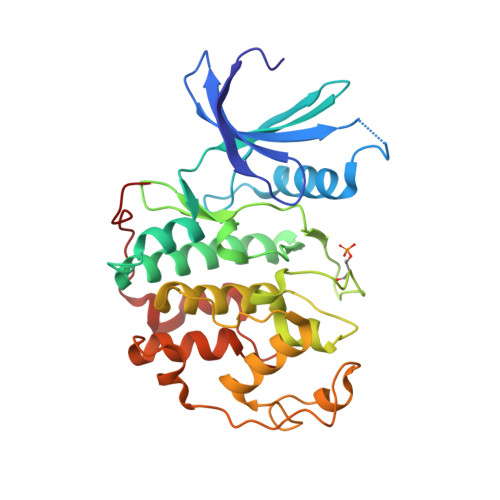
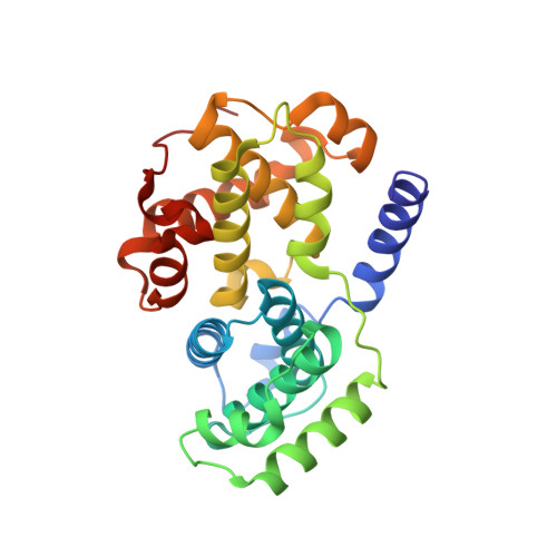
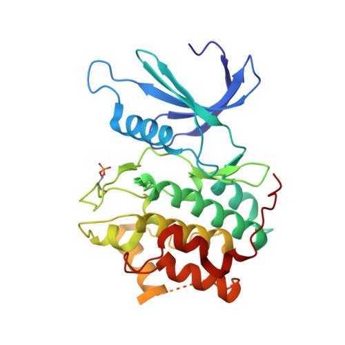
5LMK

Structure of phopsho-CDK2-cyclin A in complex with an ATP-competitive inhibitor


Experimental Data Snapshot

  • Method: X-RAY DIFFRACTION
  • Resolution: 2.40 Å
  • R-Value Free: 0.249 
  • R-Value Work: 0.212 
  • R-Value Observed: 0.213 

Starting Model: other
View more details

wwPDB Validation   3D Report Full Report


Ligand Structure Quality Assessment 


This is version 1.3 of the entry. See complete history


Literature

Explicit treatment of active-site waters enhances quantum mechanical/implicit solvent scoring: Inhibition of CDK2 by new pyrazolo[1,5-a]pyrimidines.

Hylsova, M.Carbain, B.Fanfrlik, J.Musilova, L.Haldar, S.Kopruluoglu, C.Ajani, H.Brahmkshatriya, P.S.Jorda, R.Krystof, V.Hobza, P.Echalier, A.Paruch, K.Lepsik, M.

(2016) Eur J Med Chem 126: 1118-1128

  • DOI: https://doi.org/10.1016/j.ejmech.2016.12.023
  • Primary Citation of Related Structures:  
    5LMK

  • PubMed Abstract: 

    We present comprehensive testing of solvent representation in quantum mechanics (QM)-based scoring of protein-ligand affinities. To this aim, we prepared 21 new inhibitors of cyclin-dependent kinase 2 (CDK2) with the pyrazolo[1,5-a]pyrimidine core, whose activities spanned three orders of magnitude. The crystal structure of a potent inhibitor bound to the active CDK2/cyclin A complex revealed that the biphenyl substituent at position 5 of the pyrazolo[1,5-a]pyrimidine scaffold was located in a previously unexplored pocket and that six water molecules resided in the active site. Using molecular dynamics, protein-ligand interactions and active-site water H-bond networks as well as thermodynamics were probed. Thereafter, all the inhibitors were scored by the QM approach utilizing the COSMO implicit solvent model. Such a standard treatment failed to produce a correlation with the experiment (R 2  = 0.49). However, the addition of the active-site waters resulted in significant improvement (R 2  = 0.68). The activities of the compounds could thus be interpreted by taking into account their specific noncovalent interactions with CDK2 and the active-site waters. In summary, using a combination of several experimental and theoretical approaches we demonstrate that the inclusion of explicit solvent effects enhance QM/COSMO scoring to produce a reliable structure-activity relationship with physical insights. More generally, this approach is envisioned to contribute to increased accuracy of the computational design of novel inhibitors.


  • Organizational Affiliation

    Department of Chemistry, CZ Openscreen, Masaryk University, Kamenice 5, 625 00 Brno, Czech Republic.


Macromolecules
Find similar proteins by:  (by identity cutoff)  |  3D Structure
Entity ID: 1
MoleculeChains Sequence LengthOrganismDetailsImage
Cyclin-dependent kinase 2299Homo sapiensMutation(s): 0 
Gene Names: CDK2CDKN2
EC: 2.7.11.22
UniProt & NIH Common Fund Data Resources
Find proteins for P24941 (Homo sapiens)
Explore P24941 
Go to UniProtKB:  P24941
PHAROS:  P24941
GTEx:  ENSG00000123374 
Entity Groups  
Sequence Clusters30% Identity50% Identity70% Identity90% Identity95% Identity100% Identity
UniProt GroupP24941
Sequence Annotations
Expand
  • Reference Sequence
Find similar proteins by:  (by identity cutoff)  |  3D Structure
Entity ID: 2
MoleculeChains Sequence LengthOrganismDetailsImage
Cyclin-A2
B, D
258Homo sapiensMutation(s): 0 
Gene Names: CCNA2CCN1CCNA
UniProt & NIH Common Fund Data Resources
Find proteins for P20248 (Homo sapiens)
Explore P20248 
Go to UniProtKB:  P20248
PHAROS:  P20248
GTEx:  ENSG00000145386 
Entity Groups  
Sequence Clusters30% Identity50% Identity70% Identity90% Identity95% Identity100% Identity
UniProt GroupP20248
Sequence Annotations
Expand
  • Reference Sequence
Find similar proteins by:  (by identity cutoff)  |  3D Structure
Entity ID: 3
MoleculeChains Sequence LengthOrganismDetailsImage
Cyclin-dependent kinase 2297Homo sapiensMutation(s): 0 
Gene Names: CDK2CDKN2
EC: 2.7.11.22
UniProt & NIH Common Fund Data Resources
Find proteins for P24941 (Homo sapiens)
Explore P24941 
Go to UniProtKB:  P24941
PHAROS:  P24941
GTEx:  ENSG00000123374 
Entity Groups  
Sequence Clusters30% Identity50% Identity70% Identity90% Identity95% Identity100% Identity
UniProt GroupP24941
Sequence Annotations
Expand
  • Reference Sequence
Small Molecules
Modified Residues  1 Unique
IDChains TypeFormula2D DiagramParent
TPO
Query on TPO
A
L-PEPTIDE LINKINGC4 H10 N O6 PTHR
Binding Affinity Annotations 
IDSourceBinding Affinity
6ZK BindingDB:  5LMK IC50: 37 (nM) from 1 assay(s)
Experimental Data & Validation

Experimental Data

  • Method: X-RAY DIFFRACTION
  • Resolution: 2.40 Å
  • R-Value Free: 0.249 
  • R-Value Work: 0.212 
  • R-Value Observed: 0.213 
  • Space Group: P 21 21 21
Unit Cell:
Length ( Å )Angle ( ˚ )
a = 74.466α = 90
b = 134.395β = 90
c = 149.481γ = 90
Software Package:
Software NamePurpose
REFMACrefinement
XDSdata reduction
SCALAdata scaling
PHASERphasing

Structure Validation

View Full Validation Report


Ligand Structure Quality Assessment 


Entry History 

Deposition Data

  • Released Date: 2017-01-25 
  • Deposition Author(s): Echalier, A.

Revision History  (Full details and data files)

  • Version 1.0: 2017-01-25
    Type: Initial release
  • Version 1.1: 2019-10-16
    Changes: Data collection
  • Version 1.2: 2024-05-01
    Changes: Data collection, Database references, Derived calculations, Refinement description
  • Version 1.3: 2024-10-16
    Changes: Structure summary