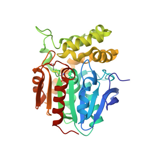
RCSB PDB - 5TNE: Crystal structure of the E153Q mutant of the CFTR inhibitory factor Cif containing the adducted cis-Stilbene Oxide hydrolysis intermediate

 5TNE

Crystal structure of the E153Q mutant of the CFTR inhibitory factor Cif containing the adducted cis-Stilbene Oxide hydrolysis intermediate

  • Classification: HYDROLASE
  • Organism(s): Pseudomonas aeruginosa UCBPP-PA14
  • Expression System: Escherichia coli
  • Mutation(s): Yes 

  • Deposited: 2016-10-14 Released: 2017-10-11 
  • Deposition Author(s): Hvorecny, K.L., Madden, D.R.
  • Funding Organization(s): Cystic Fibrosis Foundation, National Institutes of Health/National Institute of General Medical Sciences (NIH/NIGMS), National Institutes of Health/National Institute Of Allergy and Infectious Diseases (NIH/NIAID), National Institutes of Health/National Institute of Diabetes and Digestive and Kidney Disease (NIH/NIDDK), National Institutes of Health/National Institute of Environmental Health Sciences (NIH/NIEHS)

Experimental Data Snapshot

  • Method: X-RAY DIFFRACTION
  • Resolution: 1.75 Å
  • R-Value Free: 0.183 
  • R-Value Work: 0.150 
  • R-Value Observed: 0.152 

Starting Model: experimental
View more details

wwPDB Validation   3D Report Full Report


Ligand Structure Quality Assessment 

Created with Raphaël 2.3.0Worse 01 BetterLigand structure goodness of fit to experimental dataBest fitted 7F6Click on this verticalbar to view details

This is version 1.4 of the entry. See complete history


Literature

Active-Site Flexibility and Substrate Specificity in a Bacterial Virulence Factor: Crystallographic Snapshots of an Epoxide Hydrolase.

Hvorecny, K.L.Bahl, C.D.Kitamura, S.Lee, K.S.S.Hammock, B.D.Morisseau, C.Madden, D.R.

(2017) Structure 25: 697-707.e4

  • DOI: https://doi.org/10.1016/j.str.2017.03.002
  • Primary Citation of Related Structures:  
    5TND, 5TNE, 5TNF, 5TNG, 5TNH, 5TNI, 5TNJ, 5TNK, 5TNL, 5TNM, 5TNN, 5TNP, 5TNQ, 5TNR, 5TNS

  • PubMed Abstract: 

    Pseudomonas aeruginosa secretes an epoxide hydrolase with catalytic activity that triggers degradation of the cystic fibrosis transmembrane conductance regulator (CFTR) and perturbs other host defense networks. Targets of this CFTR inhibitory factor (Cif) are largely unknown, but include an epoxy-fatty acid. In this class of signaling molecules, chirality can be an important determinant of physiological output and potency. Here we explore the active-site chemistry of this two-step α/β-hydrolase and its implications for an emerging class of virulence enzymes. In combination with hydrolysis data, crystal structures of 15 trapped hydroxyalkyl-enzyme intermediates reveal the stereochemical basis of Cif's substrate specificity, as well as its regioisomeric and enantiomeric preferences. The structures also reveal distinct sets of conformational changes that enable the active site to expand dramatically in two directions, accommodating a surprising array of potential physiological epoxide targets. These new substrates may contribute to Cif's diverse effects in vivo, and thus to the success of P. aeruginosa and other pathogens during infection.


  • Organizational Affiliation

    Department of Biochemistry and Cell Biology, Geisel School of Medicine, Dartmouth College, Hanover, NH 03755, USA.


Macromolecules
Find similar proteins by:  (by identity cutoff)  |  3D Structure
Entity ID: 1
MoleculeChains Sequence LengthOrganismDetailsImage
CFTR inhibitory factor
A, B, C, D
301Pseudomonas aeruginosa UCBPP-PA14Mutation(s): 1 
Gene Names: PA14_26090
UniProt
Find proteins for A0A0H2ZD27 (Pseudomonas aeruginosa (strain UCBPP-PA14))
Explore A0A0H2ZD27 
Go to UniProtKB:  A0A0H2ZD27
Entity Groups  
Sequence Clusters30% Identity50% Identity70% Identity90% Identity95% Identity100% Identity
UniProt GroupA0A0H2ZD27
Sequence Annotations
Expand
  • Reference Sequence
Experimental Data & Validation

Experimental Data

  • Method: X-RAY DIFFRACTION
  • Resolution: 1.75 Å
  • R-Value Free: 0.183 
  • R-Value Work: 0.150 
  • R-Value Observed: 0.152 
  • Space Group: C 1 2 1
Unit Cell:
Length ( Å )Angle ( ˚ )
a = 167.944α = 90
b = 83.69β = 100.31
c = 89.175γ = 90
Software Package:
Software NamePurpose
PHENIXrefinement
XDSdata reduction
XDSdata scaling
PHENIXphasing

Structure Validation

View Full Validation Report


Ligand Structure Quality Assessment 

Created with Raphaël 2.3.0Worse 01 BetterLigand structure goodness of fit to experimental dataBest fitted 7F6Click on this verticalbar to view details

Entry History & Funding Information

Deposition Data


Funding OrganizationLocationGrant Number
Cystic Fibrosis FoundationUnited States--
National Institutes of Health/National Institute of General Medical Sciences (NIH/NIGMS)United States--
National Institutes of Health/National Institute Of Allergy and Infectious Diseases (NIH/NIAID)United States--
National Institutes of Health/National Institute of Diabetes and Digestive and Kidney Disease (NIH/NIDDK)United States--
National Institutes of Health/National Institute of Environmental Health Sciences (NIH/NIEHS)United States--

Revision History  (Full details and data files)

  • Version 1.0: 2017-10-11
    Type: Initial release
  • Version 1.1: 2019-11-06
    Changes: Author supporting evidence, Data collection
  • Version 1.2: 2019-12-11
    Changes: Author supporting evidence
  • Version 1.3: 2023-10-04
    Changes: Data collection, Database references, Refinement description
  • Version 1.4: 2024-11-06
    Changes: Structure summary