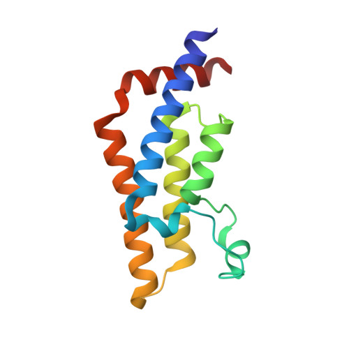
7R00

ATAD2 in complex with FragLite33


Experimental Data Snapshot

  • Method: X-RAY DIFFRACTION
  • Resolution: 1.48 Å
  • R-Value Free: 0.237 
  • R-Value Work: 0.207 

Starting Model: experimental
View more details

wwPDB Validation   3D Report Full Report


Ligand Structure Quality Assessment 


This is version 1.1 of the entry. See complete history


Literature

Mapping Ligand Interactions of Bromodomains BRD4 and ATAD2 with FragLites and PepLites─Halogenated Probes of Druglike and Peptide-like Molecular Interactions.

Davison, G.Martin, M.P.Turberville, S.Dormen, S.Heath, R.Heptinstall, A.B.Lawson, M.Miller, D.C.Ng, Y.M.Sanderson, J.N.Hope, I.Wood, D.J.Cano, C.Endicott, J.A.Hardcastle, I.R.Noble, M.E.M.Waring, M.J.

(2022) J Med Chem 65: 15416-15432

  • DOI: https://doi.org/10.1021/acs.jmedchem.2c01357
  • Primary Citation of Related Structures:  
    7PPX, 7QU7, 7QUK, 7QUM, 7QWO, 7QX1, 7QXT, 7QYK, 7QYL, 7QZM, 7QZY, 7QZZ, 7R00, 7R05, 7R0Y, 7Z9H, 7Z9I, 7Z9J, 7Z9N, 7Z9O, 7Z9S, 7Z9U, 7Z9W, 7Z9Y, 7ZA6, 7ZA7, 7ZA8, 7ZA9, 7ZAA, 7ZAD, 7ZAE, 7ZAJ, 7ZAQ, 7ZAR, 7ZAT, 7ZE6, 7ZE7, 7ZEF, 7ZEN, 7ZFN, 7ZFO, 7ZFS, 7ZFT, 7ZFU, 7ZFV, 7ZFY, 7ZFZ, 7ZG1, 7ZG2

  • PubMed Abstract: 

    The development of ligands for biological targets is critically dependent on the identification of sites on proteins that bind molecules with high affinity. A set of compounds, called FragLites, can identify such sites, along with the interactions required to gain affinity, by X-ray crystallography. We demonstrate the utility of FragLites in mapping the binding sites of bromodomain proteins BRD4 and ATAD2 and demonstrate that FragLite mapping is comparable to a full fragment screen in identifying ligand binding sites and key interactions. We extend the FragLite set with analogous compounds derived from amino acids (termed PepLites) that mimic the interactions of peptides. The output of the FragLite maps is shown to enable the development of ligands with leadlike potency. This work establishes the use of FragLite and PepLite screening at an early stage in ligand discovery allowing the rapid assessment of tractability of protein targets and informing downstream hit-finding.


  • Organizational Affiliation

    Cancer Research Horizons Therapeutic Innovation, Newcastle Drug Discovery Unit, Newcastle University Centre for Cancer, Chemistry, School of Natural and Environmental Sciences, Newcastle University, Bedson Building, Newcastle upon Tyne NE1 7RU, U.K.


Macromolecules
Find similar proteins by:  (by identity cutoff)  |  3D Structure
Entity ID: 1
MoleculeChains Sequence LengthOrganismDetailsImage
ATPase family AAA domain-containing protein 2A [auth AAA]130Homo sapiensMutation(s): 0 
Gene Names: ATAD2L16PRO2000
EC: 3.6.1.3 (PDB Primary Data), 3.6.1 (UniProt)
UniProt & NIH Common Fund Data Resources
Find proteins for Q6PL18 (Homo sapiens)
Explore Q6PL18 
Go to UniProtKB:  Q6PL18
PHAROS:  Q6PL18
GTEx:  ENSG00000156802 
Entity Groups  
Sequence Clusters30% Identity50% Identity70% Identity90% Identity95% Identity100% Identity
UniProt GroupQ6PL18
Sequence Annotations
Expand
  • Reference Sequence
Small Molecules
Ligands 4 Unique
IDChains Name / Formula / InChI Key2D Diagram3D Interactions
HWC (Subject of Investigation/LOI)
Query on HWC

Download Ideal Coordinates CCD File 
D [auth AAA]3-azanyl-5-bromanyl-1-methyl-pyridin-2-one
C6 H7 Br N2 O
KRUDZWOELKQDJW-UHFFFAOYSA-N
SO4
Query on SO4

Download Ideal Coordinates CCD File 
B [auth AAA],
K [auth AAA]
SULFATE ION
O4 S
QAOWNCQODCNURD-UHFFFAOYSA-L
EDO
Query on EDO

Download Ideal Coordinates CCD File 
E [auth AAA]
F [auth AAA]
G [auth AAA]
H [auth AAA]
I [auth AAA]
E [auth AAA],
F [auth AAA],
G [auth AAA],
H [auth AAA],
I [auth AAA],
J [auth AAA]
1,2-ETHANEDIOL
C2 H6 O2
LYCAIKOWRPUZTN-UHFFFAOYSA-N
CL
Query on CL

Download Ideal Coordinates CCD File 
C [auth AAA]CHLORIDE ION
Cl
VEXZGXHMUGYJMC-UHFFFAOYSA-M
Binding Affinity Annotations 
IDSourceBinding Affinity
HWC BindingDB:  7R00 IC50: 3.77e+5 (nM) from 1 assay(s)
Experimental Data & Validation

Experimental Data

  • Method: X-RAY DIFFRACTION
  • Resolution: 1.48 Å
  • R-Value Free: 0.237 
  • R-Value Work: 0.207 
  • Space Group: P 65 2 2
Unit Cell:
Length ( Å )Angle ( ˚ )
a = 79.331α = 90
b = 79.331β = 90
c = 137.081γ = 120
Software Package:
Software NamePurpose
REFMACrefinement
REFMACrefinement
Aimlessdata scaling
xia2data reduction
PHASERphasing

Structure Validation

View Full Validation Report


Ligand Structure Quality Assessment 


Entry History & Funding Information

Deposition Data


Funding OrganizationLocationGrant Number
Cancer Research UKUnited KingdomC57659/A27310
Cancer Research UKUnited KingdomC1362/A20263
Cancer Research UKUnited KingdomC2215/A21421

Revision History  (Full details and data files)

  • Version 1.0: 2022-12-07
    Type: Initial release
  • Version 1.1: 2024-01-31
    Changes: Data collection, Refinement description